Toggle light / dark theme

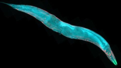
Scientists at the MDI Biological Laboratory, in collaboration with scientists from the Buck Institute for Research on Aging in Novato, Calif., and Nanjing University in China, have identified synergistic cellular pathways for longevity that amplify lifespan fivefold in C. elegans, a nematode worm used as a model in aging research.

The increase in lifespan would be the equivalent of a human living for 400 or 500 years, according to one of the scientists.

The research draws on the discovery of two major pathways governing aging in C. elegans, which is a popular model in aging research because it shares many of its genes with humans and because its short lifespan of only three to four weeks allows scientists to quickly assess the effects of genetic and environmental interventions to extend healthy lifespan.

Scientists at the MDI Biological Laboratory, in collaboration with scientists from the Buck Institute for Research on Aging in Novato, Calif., and Nanjing University in China, have identified synergistic cellular pathways for longevity that amplify lifespan fivefold in C. elegans, a nematode worm used as a model in aging research.

The increase in would be the equivalent of a human living for 400 or 500 years, according to one of the scientists.

The research draws on the discovery of two major pathways governing aging in C. elegans, which is a popular model in aging research because it shares many of its genes with humans and because its short lifespan of only three to four weeks allows scientists to quickly assess the effects of genetic and environmental interventions to extend healthy lifespan.

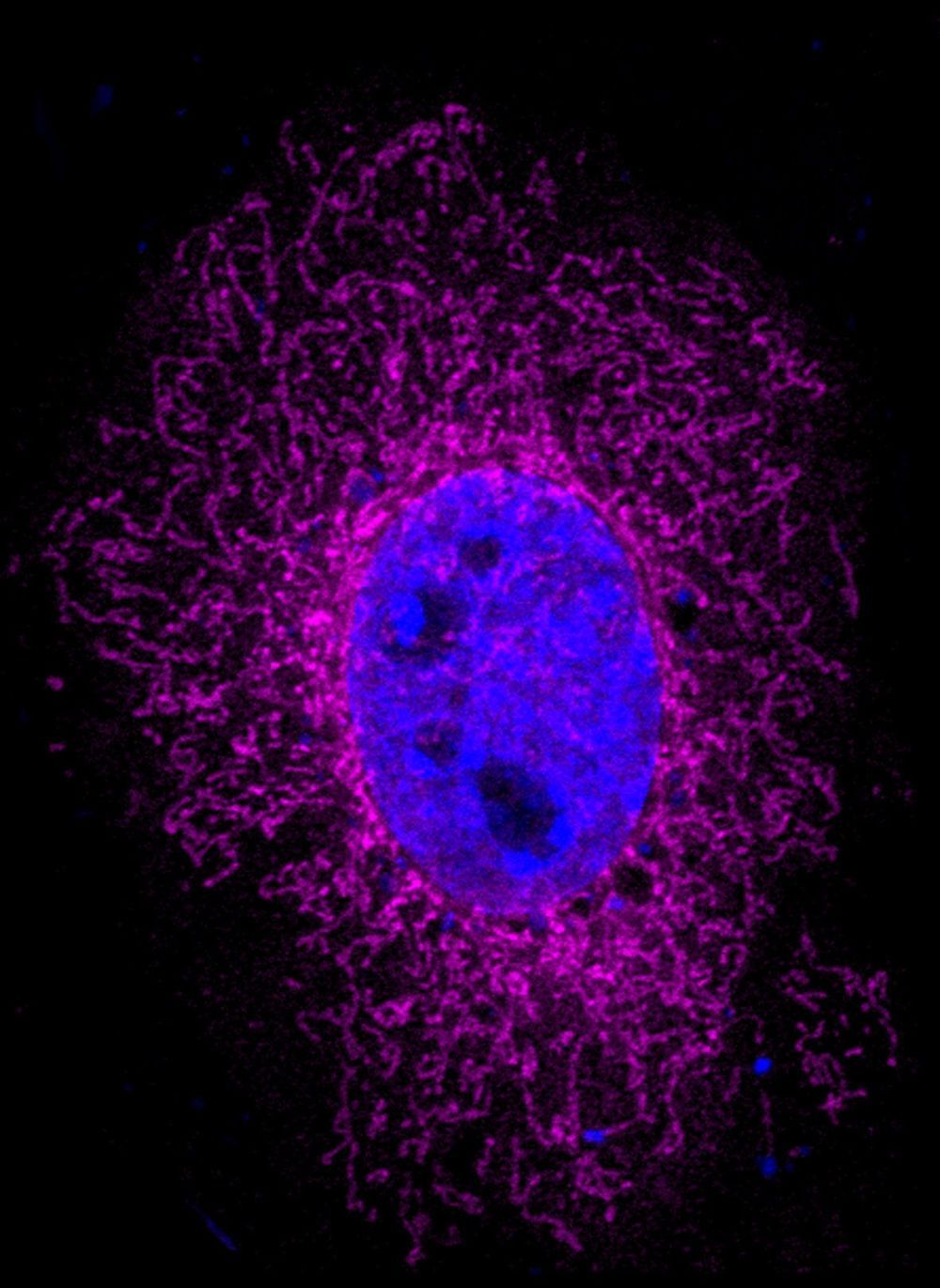
The size of the human brain increased profoundly during evolution. A certain gene that is only found in humans triggers brain stem cells to form a larger pool of stem cells. As a consequence, more neurons can arise, which paves the way to a bigger brain. This brain size gene is called ARHGAP11B and so far, how it works was completely unknown. Researchers at the Max Planck Institute of Molecular Cell Biology and Genetics in Dresden now uncovered its mode of action. They show that the ARHGAP11B protein is located in the powerhouse of the cell—the mitochondria—and induces a metabolic pathway in the brain stem cells that is characteristic of cancer cells.

The research group of Wieland Huttner, a founding director of the Max Planck Institute of Molecular Cell Biology and Genetics, has been investigating the underlying the expansion of the brain during mammalian evolution for many years. In 2015, the group reported a key role for a gene that is only present in humans and in our closest extinct relatives, the Neanderthals and Denisovans. This gene, named ARHGAP11B, causes the so-called basal brain stem to expand in number and to eventually increase the production of neurons, leading to a bigger and more folded brain in the end. How the gene functions within the basal brain stem cells has been unknown so far.

Takashi Namba, a postdoctoral scientist in the research group of Wieland Huttner, wanted to find the answer to this question, together with colleagues from the Max Planck Institute, the University Hospital Carl Gustav Carus Dresden, and the Department of Medical Biochemistry at the Semmelweis University, Budapest. He found that the ARHGAP11B protein is located in mitochondria, the organelles that generate most of the cell’s source of chemical energy and hence are often referred to as the powerhouse of the cell. Takashi Namba explains the results: We found that ARHGAP11B interacts with a protein in the membrane of mitochondria that regulates a membrane pore. As a consequence of this interaction, the pores in the membrane are closing up, preventing calcium leakage from the mitochondria. The resulting higher calcium concentration causes the mitochondria to generate chemical energy by a metabolic pathway called glutaminolysis.

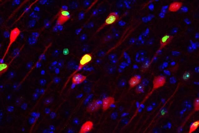
Experiments in rodents have revealed that engrams exist as multiscale networks of neurons. An experience becomes stored as a potentially retrievable memory in the brain when excited neurons in a brain region such as the hippocampus or amygdala become recruited into a local ensemble. These ensembles combine with others in other regions, such as the cortex, into an “engram complex.” Crucial to this process of linking engram cells is the ability of neurons to forge new circuit connections, via processes known as “synaptic plasticity” and “dendritic spine formation.” Importantly, experiments show that the memory initially stored across an engram complex can be retrieved by its reactivation but may also persist “silently” even when memories cannot be naturally recalled, for instance in mouse models used to study memory disorders such as early stage Alzheimer’s disease.

“More than 100 years ago Semon put forth a law of engraphy,” wrote Josselyn, Senior Scientist at SickKids, Professor of Psychology and Physiology at the University of Toronto and Senior Fellow in the Brain, Mind & Consciousness Program at the Canadian Institute for Advanced Research, (CIFAR) and Tonegawa, Picower Professor of Biology and Neuroscience at the RIKEN-MIT Laboratory for Neural Circuit Genetics at MIT and Investigator of the Howard Hughes Medical Institute. “Combining these theoretical ideas with the new tools that allow researchers to image and manipulate engrams at the level of cell ensembles facilitated many important insights into memory function.”

“For instance, evidence indicates that both increased intrinsic excitability and synaptic plasticity work hand in hand to form engrams and that these processes may also be important in memory linking, memory retrieval, and memory consolidation.”

For as much as the field has learned, Josselyn and Tonegawa wrote, there are still important unanswered questions and untapped potential applications: How do engrams change over time? How can engrams and memories be studied more directly in humans? And can applying knowledge about biological engrams inspire advances in artificial intelligence, which in turn could feedback new insights into the workings of engrams?


A review in Science traces neuroscientists’ progress in studying the neural substrate for storing memories and raises key future questions for the field.
https://www.youtube.com/watch?v=bKCZcjpzJe0&t=1s

https://www.youtube.com/watch?v=16VcRfkFeGw&t=1s&fbclid=IwAR24ZvROX7k1dOiMT51gReBdQRnMVU7KYIuF6vmASvUrWOuxTCEoVKXkcIE

Worldwide, more people die from tuberculosis (TB) than any other infectious disease, even though the vast majority were vaccinated. The vaccine just isn’t that reliable. But a new Nature study finds that simply changing the way the vaccine is administered could dramatically boost its protective power.

Researchers at the University of Pittsburgh School of Medicine and the National Institute of Allergy and Infectious Diseases (NIAID) discovered that intravenous TB vaccination is highly protective against the infection in monkeys, compared to the standard injection directly into the skin, which offers minimal protection.

“The effects are amazing,” said senior author JoAnne Flynn, Ph.D., professor of microbiology and molecular genetics at the Pitt Center for Vaccine Research. “When we compared the lungs of animals given the vaccine intravenously versus the standard route, we saw a 100,000-fold reduction in bacterial burden. Nine out of 10 animals showed no inflammation in their lungs.”

Happy New year everyone! We wanted to give you an inside look as to what happens on our Dr. Joe live calls that take place once a month. Here’s a moment from our last Dr. Joe live call.

⁣ Some of the subjects Dr. Joe may address: latest scientific discoveries; health and wealth; love and relationships; neuroscience and epigenetics; happiness and vitality; the quantum model of reality; how you can make great and lasting changes in yourself and your life!⁣

⁣ With his vast experience and deepening knowledge of how each one of us can unlock unlimited abilities and transform our lives for the better, these classes offer you a unique opportunity to continually learn and interact with Dr. Joe! If you want to take it a step further in the work, Dr. Joe live is a great way to deepen your understanding, keep you plugged into the community and interact with Dr. Joe personally.⁣.

After years of ethical debates and breakthroughs in the lab, CRISPR has finally made its way to clinical trials. Researchers are now looking at whether the DNA-editing tool, as well as more conventional gene therapies, can effectively treat a wide array of heritable disorders and even cancers.

“There’s been a convergence of the science getting better, the manufacturing getting much better, and money being available for these kinds of studies,” says Cynthia Dunbar, a senior investigator at the National Heart, Lung, and Blood Institute. “It’s truly come of age.”

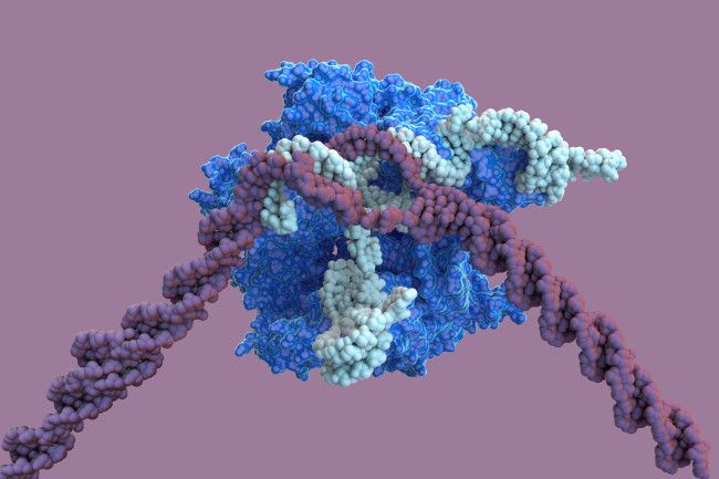
CRISPR — formally known as CRISPR-Cas9 — has been touted as an improvement over conventional gene therapy because of its potential precision. CRISPR (clustered regularly interspaced short palindromic repeats) is a genetic code that, contained in a strand of RNA and paired with the enzyme Cas9, acts like molecular scissors that can target and snip out specific genes. Add a template for a healthy gene, and CRISPR’s cut can allow the cell to replace a defective gene with a healthy one.

A group of scientists from the Russian Academy of Sciences (ICG SB RAS) and the TSU Biological Institute has established a path through which nanoparticles of viruses and organic and inorganic substances from the environment enter the brain. Additionally, the researchers report a simple and inexpensive way to block their entry. The data obtained by the project could play a large role in medicine and pharmaceuticals, where nanoparticles are increasingly used for the diagnosis and treatment of serious diseases.

“There are a large number of nanoparticles of a wide variety of chemical elements and their compounds in the environment, ranging from harmless to toxic, for example, heavy metal oxides,” says Mikhail Moshkin, director of the Center for Laboratory Animal Genetic Resources of the ICG SB RAS. “Scientists have accumulated data that indicate the adverse effect of nanoparticles, for example, people who live closer than 50 meters to large highways may develop neurodegenerative diseases (Alzheimer’s, Parkinson’s and others) due to the accumulation of nanosized particles in the brain.”

The researchers sought to determine how nanoparticles enter the brain. They cannot penetrate through the lungs and blood vessels because the blood-brain barrier blocks them from the brain. Experiments conducted on rodents helped calculate the trajectory of the movement of nanoparticles.