Toggle light / dark theme

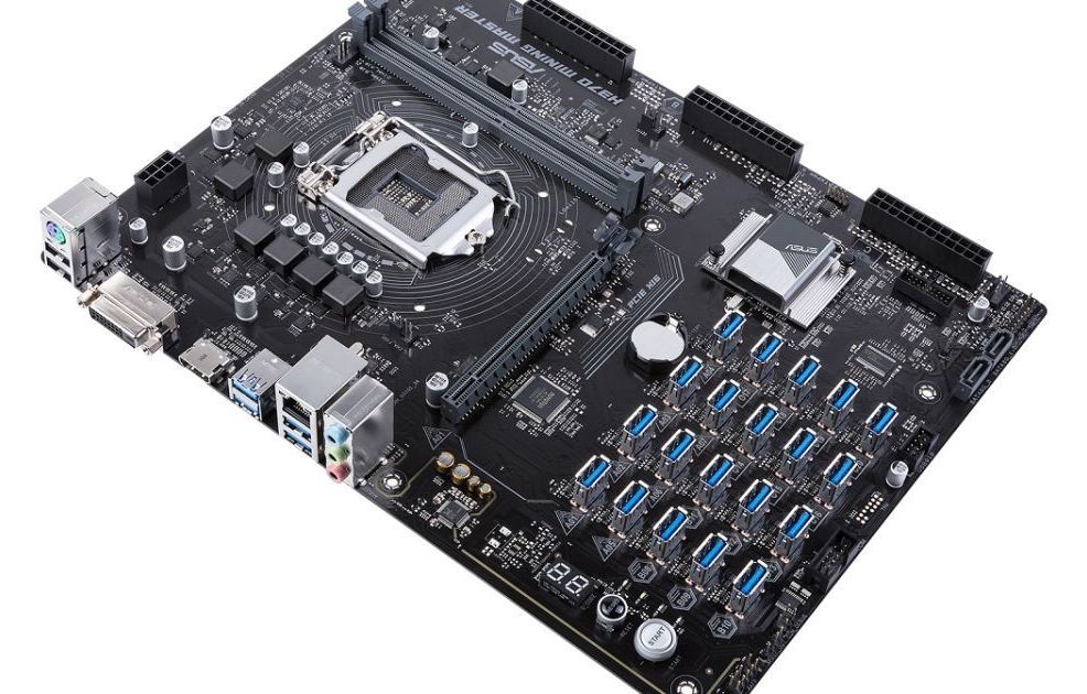
ASUS is moving further into the cryptocurrency hardware market with a motherboard that can support up to 20 graphics cards, which are typically used for mining. The H370 Mining Master uses PCIe-over-USB ports for what ASUS says is sturdier, simpler connectivity than other mining-focused motherboards.

You can manage each port and graphics card with on-board diagnostics. One feature scans your system when you boot up to determine the status of each port, while there are onboard LEDs that signify a problem with components such as memory or the processor (there’s space for an Intel 8th-gen Core CPU). ASUS has added some other features to optimize mining as well.

The H370 Mining Master follows last year’s B250 Mining Expert, which had room for 19 CPUs via PCIe ports. ASUS says that board had far more sales than it expected, which prompted the company to keep traveling down the crypto road and evolve its mining-tailored motherboards. The latest board will ship later this year, though ASUS has yet to announce pricing. You might need to fork over several Ethereum coins to buy enough graphics cards for all those spaces, though.
Read more

https://www.engadget.com/…/3D-printed-brain-medical-imagin…/


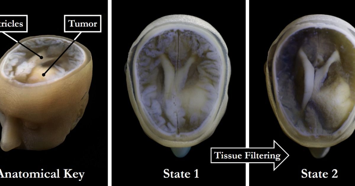
There are almost limitless possibilities when it comes to 3D printing. Design your own color-changing jewelry? Fine. Fabricate your own drugs? No problem. Print an entire house in under 24 hours? Sure! Now, researchers have come up with a fast and easy way to print palm-sized models of individual human brains, presumably in a bid to advance scientific endeavours, but also because, well, that’s pretty neat.

In theory, creating a 3D printout of a human brain has been done before, using data from MRI and CT scans. But as MIT graduate Steven Keating found when he wanted to examine his own brain following his surgery to remove a baseball-sized tumour, it’s a slow, cumbersome process that doesn’t reveal any important areas of interest.

MRI and CT scans produce images with so much detail that objects of interest need to be isolated from surrounding tissue and converted into surface meshes in order to be printed. This involves a radiologist manually tracing the desired object onto every single image “slice” of the brain, or it can be done via automatic thresholding, where a computer converts areas that contain grayscale pixels into either solid black or solid white pixels, based on a shade of gray that is chosen to be the threshold between black and white. But since medical imaging data often contains irregularly-shaped objects and lacks clear borders, features of interest are usually over- or under-exaggerated, and details are washed out.

Read more

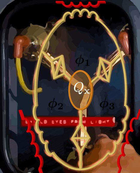
Physicists have invented a flux capacitor and, while it might not run a ‘Back to the Future’ inspired time machine, they say it will have important applications in communication technology and quantum computing.

The team from The University of Queensland, RMIT University and ETH Zurich have proposed a device which uses the quantum tunnelling of magnetic flux around a capacitor which they say can break time-reversal symmetry.

UQ Professor Tom Stace said the research proposed a new generation of electronic circulators – devices that control the direction in which microwave signals move.

Read more

Swedes have gone on to be very active in microchipping, with scant debate about issues surrounding its use, in a country keen on new technology and where the sharing of personal information is held up as a sign of a transparent society.

Ms Ulrika Celsing, 28, is one of 3,000 Swedes to have injected a microchip into her hand to try out a new way of life.

To enter her workplace, the media agency Mindshare, she simply waves her hand on a small box and types in a code before the doors open.

Read more

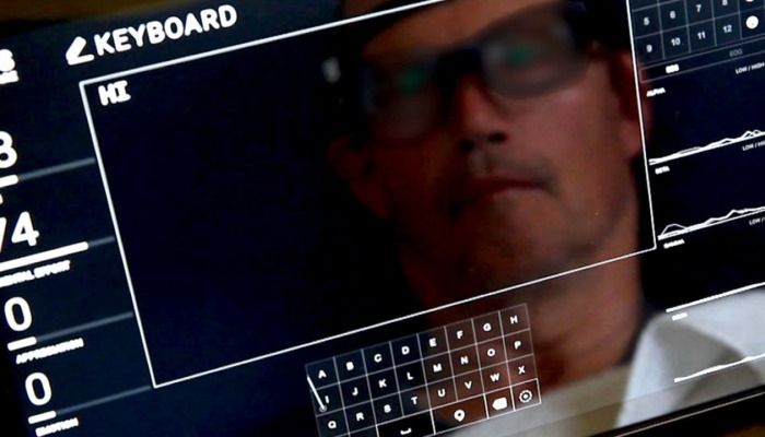
Tesla’s Elon Musk and Facebook’s Mark Zuckerberg each aim to create the world’s first brain-computer interface, but a start-up called Nuro could beat them to the punch with a simpler piece of software.

Business Insider UPDATED : Friday, 25 May 2018, 3:02PM

Read more

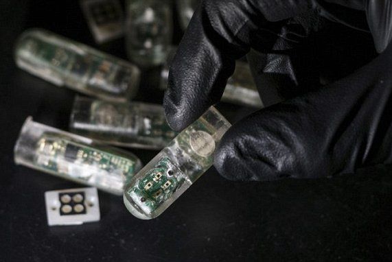
Unlike other ingestible chips, this new version by MIT contains millions of genetically engineered living cells to act as sensors, designed to light up when they detect bleeding.

It’s the latest advance in a growing field of sensors that can be swallowed or worn to monitor our health.

Pills equipped with cameras, thermometers and acidity gauges already look for disease and track digestion.

Read more

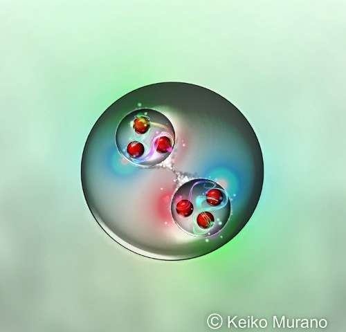
Based on complex simulations of quantum chromodynamics performed using the K computer, one of the most powerful computers in the world, the HAL QCD Collaboration, made up of scientists from the RIKEN Nishina Center for Accelerator-based Science and the RIKEN Interdisciplinary Theoretical and Mathematical Sciences (iTHEMS) program, together with colleagues from a number of universities, have predicted a new type of “dibaryon”—a particle that contains six quarks instead of the usual three. Studying how these elements form could help scientists understand the interactions among elementary particles in extreme environments such as the interiors of neutron stars or the early universe moments after the Big Bang.

Particles known as “baryons”—principally protons and neutrons—are composed of three quarks bound tightly together, with their charge depending on the “color” of the quarks that make them up. A dibaryon is essentially a system with two baryons. There is one known dibaryon in nature—deuteron, a deuterium (or heavy-hydrogen) nucleus that contains a proton and a that are very lightly bound. Scientists have long wondered whether there could be other types of dibaryons. Despite searches, no other dibaryon has been found.

The group, in work published in Physical Review Letters, has now used powerful theoretical and computational tools to predict the existence of a “most strange” dibaryon, made up of two “Omega baryons” that contain three strange quarks each. They named it “di-Omega”. The group also suggested a way to look for these strange through experiments with heavy ion collisions planned in Europe and Japan.

Read more

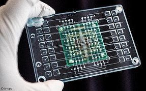
Imec, the world-leading research and innovation hub in nano-electronics and digital technologies, presents this week at its technology forum ITF 2018 (Antwerp, May 23–24), a novel organ-on-chip platform for pharmacological studies with unprecedented signal quality. It fuses imec’s high-density multi-electrode array (MEA)-chip with a microfluidic well plate, developed in collaboration with Micronit Microtechnologies, in which cells can be cultured, providing an environment that mimics human physiology. Capable of performing multiple tests in parallel, the new device aims to be a game-changer for the pharmaceutical industry, offering high quality data in the drug development process.

Every year a handful of new drugs make it to the market, but in their wake tens of thousands of candidate drugs didn’t make the cut. Nevertheless, this journey will have taken a decade and costs billions. The fact that is so time-consuming and costly, is because of the insufficiency of the existing methodologies for screening assays. These current assays are based on poor cell models that limit the quality of the resulting data, and result in inadequate biological relevance. Additionally, there is a lack of spatial resolution of the assays, resulting in the inability to screen single cells in a cell culture. Imec’s novel organ-on-chip platform aims to address these shortcomings and challenges.

Imec’s solution packs 16,384 electrodes, distributed over 16 wells, and offers multiparametric analysis. Each of the 1,024 electrodes in a well can detect intracellular action potentials, aside from the traditional extracellular signals. Further, ’s chip is patterned with microstructures to allow for a structured cell growth mimicking a specific organ.

Read more

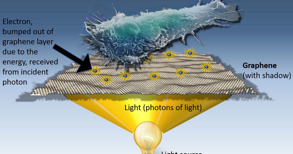
A chance lab discovery is opening up the possibility for wide-scale improvements in drug screening, application of selective painkillers, and selectively nuking cancer cells. The mystery material? Graphene, a semi-metal that’s composed of a single layer of carbon atoms. It’s already being used to make flexible OLED displays and reduce the energy costs of desalination, but its potential benefits for the medical field look promising too.

It began with a theory — scientists at the University of California knew graphene could convert light into electricity, and wondered whether that electricity had the capacity to stimulate human cells. Graphene is extremely sensitive to light (1,000 times more than traditional digital cameras and smartphones) and after experimenting with different light intensities, Alex Savchenko and his team discovered that cells could indeed be stimulated via optical graphene stimulation.

“I was looking at the microscope’s computer screen and I’m turning the knob for light intensity and I see the cells start beating faster,” he said. “I showed that to our grad students and they were yelling and jumping and asking if they could turn the knob. We had never seen this possibility of controlling cell contraction.”

Read more