Toggle light / dark theme

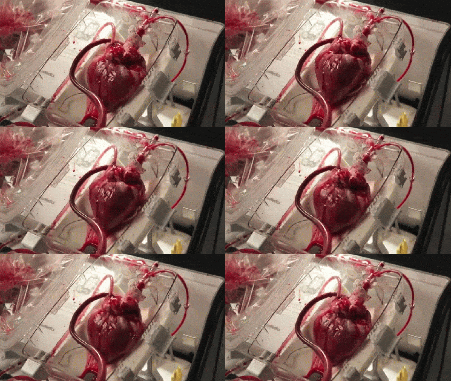
A device that reanimates organs taken from dead patients has shown promise in heart transplant surgeries, though it’s raising some ethical concerns, as well. As MIT Technology Review reports, the so-called “heart in a box” uses tubing and oxygen to pump blood and electrolytes into hearts from recently deceased patients, allowing the organs to continue functioning within a chamber. The system, developed by Massachusetts-based Transmedics, has been successfully deployed in at least 15 heart transplants in the UK and Australia, and is awaiting regulatory approval in the US.

Until now, hearts used for transplants have usually been extracted from brain-dead patients; those from dead patients have been considered too damaged. Once removed, the hearts are also stored and transported in cold temperatures to avoid rapid deterioration, though scientists have begun using devices like the heart in a box to keep the organs warm and functioning. That, doctors say, could increase the pool of donated hearts by between 15 and 30 percent.

Some say the $250,000 device is still too expensive to be deployed widely, and that it needs greater automation. For medical ethicists, the question is how long surgeons should wait before removing a heart that has stopped. “How can you say it’s irreversible, when the circulatory function is restored in a different body?” Robert Truog, an ethicist at Harvard University, tells MIT Technology Review. “We tend to overlook that because we want to transplant these organs.” Truog says he believes those patients can be considered dead, though it’s ultimately a decision for family members to make. “They are dying and it’s permissible to use their organs. The question is whether they are being harmed, and I would say they are not.”
Read more

A key mystery of the DNA replication process has been unraveled by researchers from King Abdullah University of Science and Technology (KAUST).

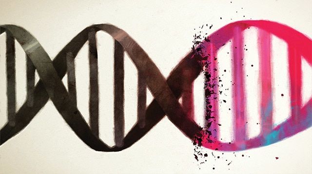
Before a bacterium can divide, it must make a copy of its genetic material, the circular DNA molecules that resemble bunched rubber bands, through a process called DNA replication. In this process, the two strands of DNA making up the circular DNA molecule unwind and separate to become templates for generating new strands.

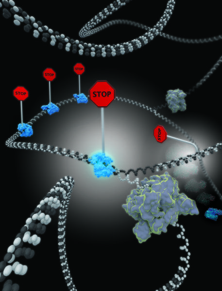
To ensure the process is well regulated, the bacterium has set a number of “roadblocks,” or termination sites on the DNA, to ensure the permanent stoppage of replication forks, Y-shaped structures formed between the strands as the DNA molecule splits.

Read more

A UCSF-led team has developed a technique to build tiny models of human tissues, called organoids, more precisely than ever before using a process that turns human cells into a biological equivalent of LEGO bricks. These mini-tissues in a dish can be used to study how particular structural features of tissue affect normal growth or go awry in cancer. They could be used for therapeutic drug screening and to help teach researchers how to grow whole human organs.

The new technique — called DNA Programmed Assembly of Cells (DPAC) and reported in the journal Nature Methods on August 31, 2015 — allows researchers to create arrays of thousands of custom-designed organoids, such as models of human mammary glands containing several hundred cells each, which can be built in a matter of hours.

There are few limits to the tissues this technology can mimic, said Zev Gartner, PhD, the paper’s senior author and an associate professor of pharmaceutical chemistry at UCSF. “We can take any cell type we want and program just where it goes. We can precisely control who’s talking to whom and who’s touching whom at the earliest stages. The cells then follow these initially programmed spatial cues to interact, move around, and develop into tissues over time.”

Read more

Engineers at the University of Toronto just made assembling functional heart tissue as easy as fastening your shoes. The team has created a biocompatible scaffold that allows sheets of beating heart cells to snap together just like Velcro™.

“One of the main advantages is the ease of use,” says biomedical engineer Professor Milica Radisic, who led the project. “We can build larger tissue structures immediately before they are needed, and disassemble them just as easily. I don’t know of any other technique that gives this ability.”

Growing heart muscle cells in the lab is nothing new. The problem is that too often, these cells don’t resemble those found in the body. Real heart cells grow in an environment replete with protein scaffolds and support cells that help shape them into long, lean beating machines. In contrast, lab-grown cells often lack these supports, and tend to be amorphous and weak. Radisic and her team focus on engineering artificial environments that more closely imitate what cells see in the body, resulting in tougher, more robust cells.

Read more

What would you say if I told you that aging happens not because of accumulation of stresses, but rather because of the intrinsic properties of the gene network of the organism? I’m guessing you’d be like: :o.

So, here’s the deal. My biohacker friends led by Peter Fedichev and Sergey Filonov in collaboration with my old friend and the longevity record holder Robert Shmookler Reis published a very cool paper. They proposed a way to quantitatively describe the two types of aging – negligible senescence and normal aging. We all know that some animals just don’t care about time passing by. Their mortality doesn’t increase with age. Such negligibly senescent species include the notorious naked mole rat and a bunch of other critters like certain turtles and clams to name a few. So the paper explains what it is exactly that makes these animals age so slowly – it’s the stability of their gene networks.

What does network stability mean then? Well, it’s actually pretty straightforward – if the DNA repair mechanisms are very efficient and the connectivity of the network is low enough, then this network is stable. So, normally aging species, such as ourselves, have unstable networks. This is a major bummer by all means. But! There is a way to overcome this problem, according to the proposed math model.

The model very generally describes what happens with a gene network over time – the majority of the genes are actually working perfectly, but a small number doesn’t. There are repair mechanisms that take care of that. Also, there are mechanisms that take care of defected proteins like heat shock proteins, etc. Put together all of this in an equasion and solve it, and bam! here’s an equasion that gives you the Gompertz law for all species that have normal aging, and a time independent constant for the negligibly senescent ones.

What’s the difference between those two aging regimes? The model suggests it’s the right combination of DNA repair efficiency and the combined efficiency of proteolysis and heat shock response systems, mediating degradation and refolding of misfolded proteins. So, it’s not the the accumulation of damages that is responsible for aging, but rather the properties of the gene network itself. The good news is that even we are playing with a terrible hand at first, there is a chance we can still win by changing the features of our network and making it stable. For example, by optimizing misfolded protein response or DNA repair.

Read more

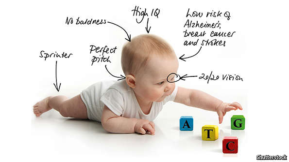
THE genome is written in an alphabet of just four letters. Being able to read, study and compare DNA sequences for humans, and thousands of other species, has become routine. A new technology promises to make it possible to edit genetic information quickly and cheaply. This could correct terrible genetic defects that blight lives. It also heralds the distant prospect of parents building their children to order.

The technology is known as CRISPR-Cas9, or just CRISPR. It involves a piece of RNA, a chemical messenger, designed to target a section of DNA; and an enzyme, called a nuclease, that can snip unwanted genes out and paste new ones in. Other ways of editing DNA exist, but CRISPR holds the promise of doing so with unprecedented simplicity, speed and precision.

Read more

Dr DePinho released a paper in 2012, this builds on previous papers and his theory of the “telomere-p53-PGC axis”. This is a big reason along with the work of Dr Michael Fossel I believe telomerase therapy is probably the best chance of radical life extension in the near future. This is one of a number of papers that implicate dysfunctional telomeres in a cascade that causes mitochondrial dysfunction and various other aging consequences.

ABSTRACT Telomere dysfunction activates p53-mediated cellular growth arrest, senescence and apoptosis to drive progressive atrophy and functional decline in high-turnover tissues. The broader adverse impact of telomere dysfunction across many tissues including more quiescent systems prompted transcriptomic network analyses to identify common mechanisms operative in haematopoietic stem cells, heart and liver. These unbiased studies revealed profound repression of peroxisome proliferator-activated receptor gamma, coactivator 1 alpha and beta (PGC-1α and PGC-1β, also known as Ppargc1a and Ppargc1b, respectively) and the downstream network in mice null for either telomerase reverse transcriptase (Tert) or telomerase RNA component (Terc) genes. Consistent with PGCs as master regulators of mitochondrial physiology and metabolism, telomere dysfunction is associated with impaired mitochondrial biogenesis and function, decreased gluconeogenesis, cardiomyopathy, and increased reactive oxygen species. In the setting of telomere dysfunction, enforced Tert or PGC-1α expression or germline deletion of p53 (also known as Trp53) substantially restores PGC network expression, mitochondrial respiration, cardiac function and gluconeogenesis. We demonstrate that telomere dysfunction activates p53 which in turn binds and represses PGC-1α and PGC-1β promoters, thereby forging a direct link between telomere and mitochondrial biology. We propose that this telomere-p53-PGC axis contributes to organ and metabolic failure and to diminishing organismal fitness in the setting of telomere dysfunction.

Read more

Cancer researchers dream of the day they can force tumor cells to morph back to the normal cells they once were. Now, researchers on Mayo Clinic’s Florida campus have discovered a way to potentially reprogram cancer cells back to normalcy.

The finding, published in Nature Cell Biology, represents “an unexpected new biology that provides the code, the software for turning off cancer,” says the study’s senior investigator, Panos Anastasiadis, Ph.D., chair of the Department of Cancer Biology on Mayo Clinic’s Florida campus.

That code was unraveled by the discovery that adhesion proteins — the glue that keeps cells together — interact with the microprocessor, a key player in the production of molecules called microRNAs (miRNAs). The miRNAs orchestrate whole cellular programs by simultaneously regulating expression of a group of genes. The investigators found that when normal cells come in contact with each other, a specific subset of miRNAs suppresses genes that promote cell growth. However, when adhesion is disrupted in cancer cells, these miRNAs are misregulated and cells grow out of control. The investigators showed, in laboratory experiments, that restoring the normal miRNA levels in cancer cells can reverse that aberrant cell growth.

Read more