Experts at Weill Cornell Medicine in New York are using gene editing tool CRISPR to alter a string of human genetic code which is known to increase the risk of developing some cancers.


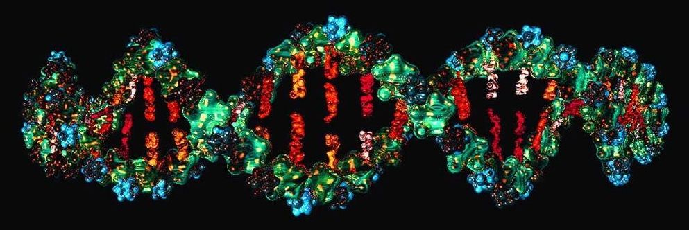
Gene editing can turn living cells into minicomputers that can read, write and perform complex calculations. The technology could track what happens inside the body over time.
DNA computers have been around since the 1990s, when researchers created DNA molecules able to perform basic mathematical functions. Instead of storing information as 0s and 1s like digital computers do, these computers store information in the molecules A, C, G and T that make up DNA.

An international team of researchers with partial support from the National Institute of Biomedical Imaging and Bioengineering (NIBIB) developed a new MRI technique that can capture an image of a brain thinking by measuring changes in tissue stiffness. The results show that brain function can be tracked on a time scale of 100 milliseconds – 60 times faster than previous methods. The technique could shed new light on altered neuronal activity in brain diseases.
The human brain responds almost immediately to stimuli, but non-invasive imaging techniques haven’t been able to keep pace with the brain. Currently, several non-invasive brain imaging methods measure brain function, but they all have limitations. Most commonly, clinicians and researchers use functional magnetic resonance imaging (fMRI) to measure brain activity via fluctuations in blood oxygen levels. However, a lot of vital brain activity information is lost using fMRI because blood oxygen levels take about six seconds to respond to a stimulus.
Since the mid-1990s, researchers have been able to generate maps of tissue stiffness using an MRI scanner, with a non-invasive technique called magnetic resonance elastography (MRE). Tissue stiffness can’t be measured directly, so instead researchers use MRE to measure the speed at which mechanical vibrations travel through tissue. Vibrations move faster through stiffer tissues, while vibrations travel through softer tissue more slowly; therefore, tissue stiffness can be determined. MRE is most commonly used to detect the hardening of liver tissue but has more recently been applied to other tissues like the brain.
Circa 2017
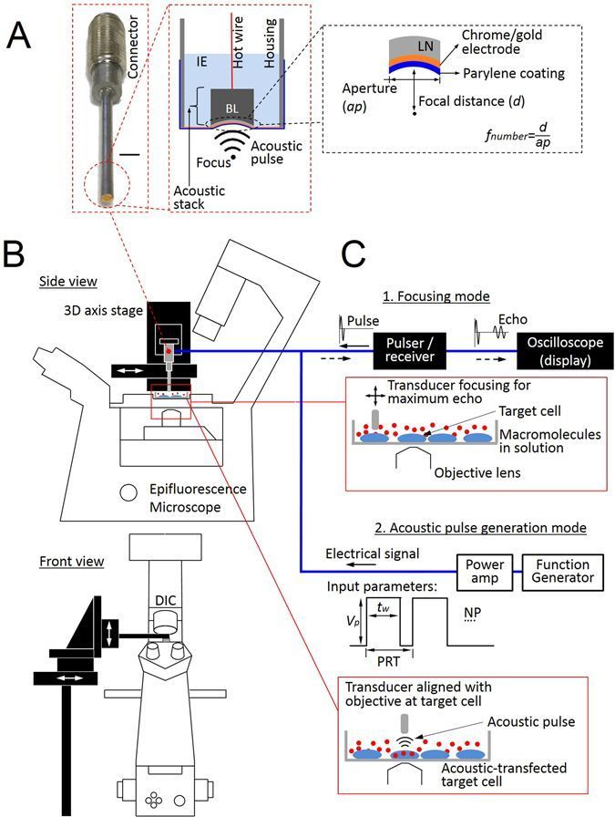
Efficient intracellular delivery of biologically active macromolecules has been a challenging but important process for manipulating live cells for research and therapeutic purposes. There have been limited transfection techniques that can deliver multiple types of active molecules simultaneously into single-cells as well as different types of molecules into physically connected individual neighboring cells separately with high precision and low cytotoxicity. Here, a high frequency ultrasound-based remote intracellular delivery technique capable of delivery of multiple DNA plasmids, messenger RNAs, and recombinant proteins is developed to allow high spatiotemporal visualization and analysis of gene and protein expressions as well as single-cell gene editing using clustered regularly interspaced short palindromic repeats (CRISPR)-associated protein-9 nuclease (Cas9), a method called acoustic-transfection. Acoustic-transfection has advantages over typical sonoporation because acoustic-transfection utilizing ultra-high frequency ultrasound over 150 MHz can directly deliver gene and proteins into cytoplasm without microbubbles, which enables controlled and local intracellular delivery to acoustic-transfection technique. Acoustic-transfection was further demonstrated to deliver CRISPR-Cas9 systems to successfully modify and reprogram the genome of single live cells, providing the evidence of the acoustic-transfection technique for precise genome editing using CRISPR-Cas9.

From computer hacking to biohacking, Dave Asprey has embarked on a quest to reverse the aging process.
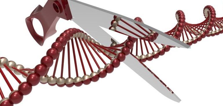
A research group at ETH Zurich, Switzerland, has made it possible to edit hundreds of genes at once with CRISPR gene editing.
CRISPR gene editing has revolutionized the biotech industry by providing an easy and quick way to genetically modify organisms. So far, however, CRISPR techniques have only managed to edit a maximum of seven genes at once. This limits the potential of the technique in creating cell therapies, since whole networks of genes need to be reprogrammed to control each cell’s fate.
The Swiss research group devised a way to overcome this limitation with a CRISPR technique able to edit 25 genes in one go. This number could also be increased to up to hundreds of genes at a time. This method therefore makes it possible to edit gene networks, and reprogram stem cells to become cell therapies such as skin cells or insulin-producing pancreatic cells.
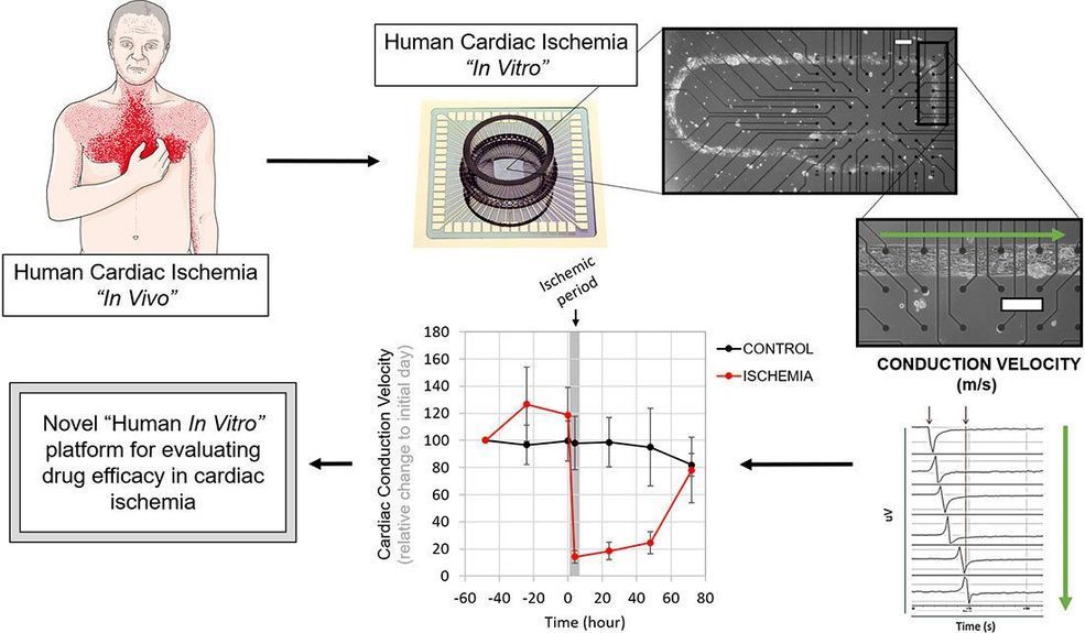
Animal models provide benefits for biomedical research, but translating such findings to human physiology can be difficult. The human heart’s energy needs and functions are difficult to reproduce in other animals, such as mice and rats. One new system looks to circumvent these issues and provide a functional view of how different treatments can help ailing cells in the heart following oxygen and nutrient deprivations.
Researchers have unveiled a new silicon chip that holds human lab-grown heart muscle cells for assessing the effectiveness of new drugs. The system includes heart cells, called cardiomyocytes, patterned on the chip with electrodes that can both stimulate and measure electrical activity within the cells. The researchers discuss their work in this week’s APL Bioengineering.
These capabilities provide a way for determining how the restriction of blood supply, a dangerous state known as ischemia, changes a heart’s conduction velocity, beat frequency and important electrical intervals associated with heart function.