Toggle light / dark theme

ReVector researchers have expertise in synthetic biology, human microbiome, and mosquito studies.


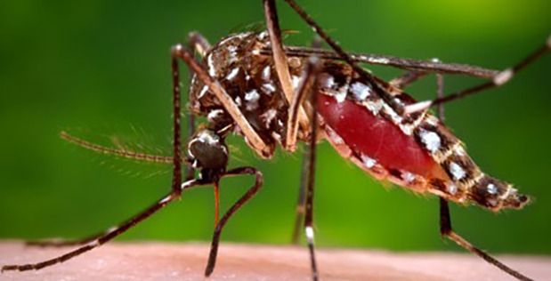
The American Society for Microbiology estimates that there are trillions of microbes living in or on the human body that constitute the human microbiome1. The human skin microbiome (HSM) acts as a barrier between humans and our external environment, protecting us from infection, but also potentially producing molecules that attract mosquitos. Mosquitos are of particular concern to the Department of Defense, as they transmit pathogens that cause diseases such as chikungunya, Zika, dengue, West Nile virus, yellow fever, and malaria. The ReVector program aims to maintain the health of military personnel operating in disease-endemic regions by reducing attraction and feeding by mosquitos, and limiting exposure to mosquito-transmitted diseases.

Genome engineering has progressed to the point where editing the HSM to remove the molecules that attract mosquitos or add genes that produce mild mosquito repellants are now possible. While the skin microbiome has naturally evolved to modulate our interactions with the environment and organisms that surround us, exerting precise control over our microbiomes is an exciting new way to provide protection from mosquito-borne diseases.

In order to advance that concept, DARPA has awarded ReVector Phase 1 contracts to two organizations: Stanford University and Ginkgo Bioworks. These performers are tasked with developing precise, safe, and efficacious technologies to modulate the profile of skin-associated volatile molecules by altering the organisms that are present in the skin microbiome and/or their metabolic processes.
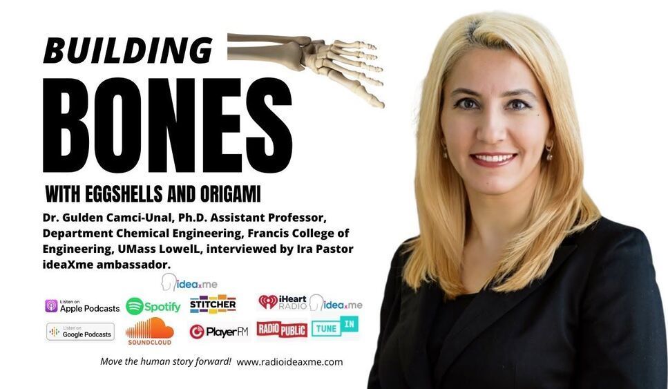
Origami-inspired tissue engineering — using eggshells, plant leaves, marine sponges, and paper as substrates.


Ira Pastor ideaXme life sciences ambassador interviews Dr. Gulden Camci-Unal, Ph.D. Assistant Professor, at the Department Chemical Engineering, Francis College of Engineering, UMass Lowell.

Ira Pastor comments:

A few episodes ago ideaXme hosted the University of Michigan’s Dr. Bruce Carlson. We spoke to him about the interesting topic of the importance of “substrate” in regenerative processes, for both the maintenance of normal tissue functions, and in the migration of cells or changes to tissue architecture that are part of healing processes.

Substrate is broadly defined as the surface or material on, or from which, cells / tissues live, grow, or obtain nourishment, and have both biochemical, as well as biomechanical functions.

Today, on ideaXme to discuss some really fascinating, next generation work that is going on in this domain, we are joined Dr. Gulden Camci-Unal, Ph.D. Assistant Professor, Department Chemical Engineering, Francis College of Engineering, UMass Lowell.

Dr Camci-Unal received her Ph.D. in Chemistry at Iowa State University (USA) and her M.Sc. and B.Sc. degrees both in Chemical Engineering at Middle East Technical University (Turkey).

Dr. Camci-Unal’s research is at the interface of biomaterials and bioengineering, including the design, synthesis, and characterization of functional biomaterials for applications in tissue engineering and regenerative medicine, development of in vitro disease models for personalized medicine, as well as work in the area low-cost point of care diagnostics.

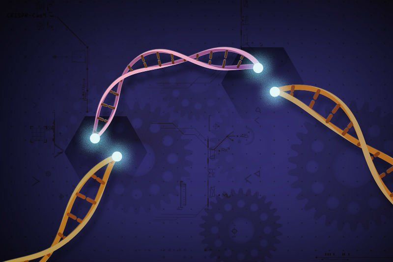
PerkinElmer has moved to expand its life sciences portfolio with CRISPR and gene editing offerings by snapping up the cell engineering specialist Horizon Discovery.

The $383 million, all-cash deal will add gene modulation tools that—in combination with its own work in applied genomics solutions—aims to provide next-generation research tools and the customized cell lines necessary for developers of new targeted therapies, and broaden PerkinElmer’s partnership work with academic researchers and the biopharma industry.

The Cambridge, U.K.-based Horizon, with about 400 employees worldwide with offices in the U.S. and Japan, provides genetic base editing technologies for living cell models using CRISPR reagents, as well as gene modulation products using RNA interference methods.

Three actions policymakers and business leaders can take today.


New developments in AI could spur a massive democratization of access to services and work opportunities, improving the lives of millions of people around the world and creating new commercial opportunities for businesses. Yet they also raise the specter of potential new social divides and biases, sparking a public backlash and regulatory risk for businesses. For the U.S. and other advanced economies, which are increasingly fractured along income, racial, gender, and regional lines, these questions of equality are taking on a new urgency. Will advances in AI usher in an era of greater inclusiveness, increased fairness, and widening access to healthcare, education, and other public services? Or will they instead lead to new inequalities, new biases, and new exclusions?

Three frontier developments stand out in terms of both their promised rewards and their potential risks to equality. These are human augmentation, sensory AI, and geographic AI.

Human Augmentation

Variously described as biohacking or Human 2.0, human augmentation technologies have the potential to enhance human performance for good or ill.

Human body bio-factories of tommorow for organ and tissue replacement.


Ira Pastor, ideaXme life sciences ambassador interviews Dr Alexander Titus Chief Strategy Officer (CSO) at the Advanced Regenerative Manufacturing Institute (ARMI).

Ira Pastor comments:

The Advanced Regenerative Manufacturing Institute (ARMI) is one of 14 institutes of the Manufacturing USA network, and is a member-driven, non-profit organization, whose mission is to make practical the large-scale manufacturing of engineered tissues and tissue-related technologies.

BioFabUSA, created by ARMI, was established to lead the charge in large-scale manufacturing of engineered tissues and regenerative medicine research, turning foundational breakthroughs in the manufacture of engineered tissues and tissue-related technologies into life-changing possibilities for everyone.

Dr. Alexander Titus is the Chief Strategy Officer (CSO) at the Advanced Regenerative Manufacturing Institute (ARMI) where he is part of the leadership team working to advance the U.S. regenerative manufacturing industry, as well as develop technologies for disaster preparedness. Dr. Titus’s career is focused on the intersection of technology and public benefit, with experience spanning the private and public sectors, as well as non-profits and academia.

Prior to his role ARMI, Dr Titus was the inaugural Assistant Director for Biotechnology, within the Office of the CTO at the Department of Defense (DoD), where he was the Deputy Assistant Secretary of Defense-level senior executive in charge of the DoD’s enterprise strategy for biotechnology, where he led the team developing the biotechnology modernization roadmap for the DoD.

Dr. Titus joined the DoD from McKinsey & Company, where he was a management consultant and a member of the inaugural cohort of Defense & Security Specialists working with the national security community on high-priority issues related to organization effectiveness, leadership, and analytics.

One of the most important questions in science is how life began on Earth.

One theory is that wet-dry cycling on the early Earth—whether through rainy/dry periods, or through phenomena such as geysers—encouraged molecular complexity. The hydration/rehydration cycle is thought to have created conditions that allowed membraneless compartments called complex coacervates to act as homes for chemicals to combine to create life.

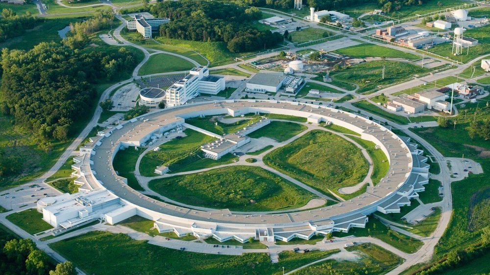
Using the Advanced Photon Source at Argonne National Laboratory, scientists in the Pritzker School of Molecular Engineering (PME) at the University of Chicago studied these compartments as they undergo phase changes to understand just what happens inside them during wet-dry cycle.

KENNEDY SPACE CENTER (FL), October 19, 2020 – The Center for the Advancement of Science in Space (CASIS) and the National Science Foundation (NSF) announced three flight projects that were selected as part of a joint solicitation focused on leveraging the International Space Station (ISS) U.S. National Laboratory to further knowledge in the fields of tissue engineering and mechanobiology. Through this collaboration, CASIS, manager of the ISS National Lab, will facilitate hardware implementation, in-orbit access, and astronaut crew time on the orbiting laboratory. NSF invested $1.2 million in the selected projects, which are seeking to advance fundamental science and engineering knowledge for the benefit of life on Earth.

This is the third collaborative research opportunity between CASIS and NSF focused on tissue engineering. Fundamental science is a major line of business for the ISS National Lab, and by conducting research in the persistent microgravity environment offered by the orbiting laboratory, NSF and the ISS National Lab will drive new advances that will bring value to our nation and spur future inquiries in low Earth orbit.

Microgravity affects organisms—from viruses and bacteria to humans, inducing changes such as altered gene expression and DNA regulation, changes in cellular function and physiology, and 3D aggregation of cells. Spaceflight is advancing research in the fields of pharmaceutical research, disease modeling, regenerative medicine, and many other areas within the life sciences. The selected projects will utilize the ISS National Lab and its unique environment to advance fundamental and transformative research that integrates engineering and life sciences.

“This is kind of a nice bookend to 16 years of research,” says Deisseroth, a neuroscientist and bioengineer at Stanford University. “It took years and years for us to sort out how to make it work.”

“The result is described this month in the journal Nature Biotechnology.”

“Optogenetics involves genetically engineering animal brains to express light-sensitive proteins—called opsins—in the membranes of neurons.”


Optogenetics can now control neural circuits at unprecedented depths within living brain tissue without surgery.

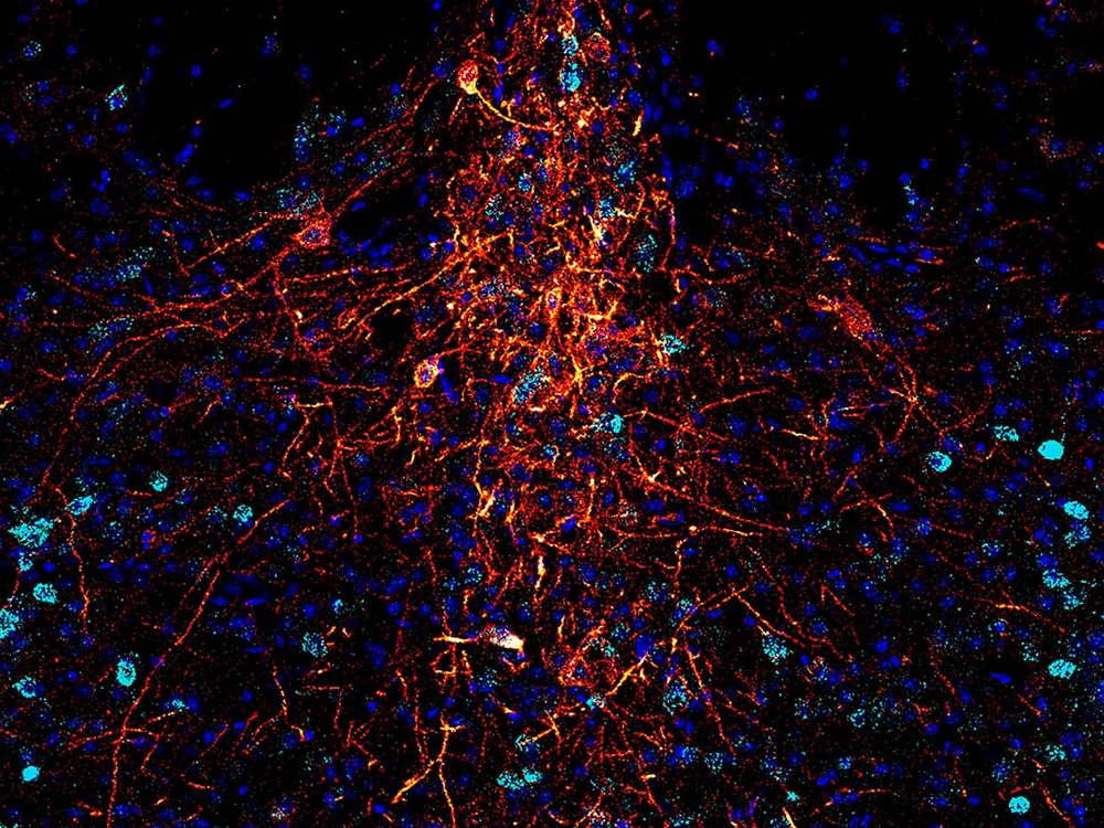
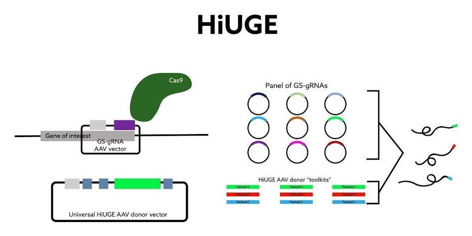
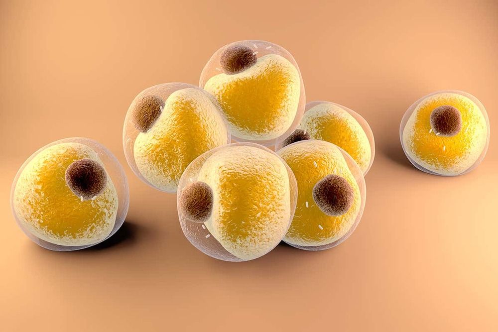
A team of researchers at Duke University have developed an imaging technology for tagging structures at a cellular level that overcomes the shortcomings of existing antibody-based techniques. Immunofluorescence imaging is a key part of the cell biologist’s toolbox, in which a fluorescent ‘flare’ attached to an antibody allows them to visualize the presence of specific target proteins in cell or tissue samples. The issue is that this specificity isn’t always 100 percent — sometimes the antibodies bind to other closely related proteins as well, making it difficult to interpret the results.

Duke’s cell biology chair Scott Soderling has led a team that developed Homology-independent Universal Genome Engineering (HiUGE), an innovation that uses gene-editing technology to rise above the shortcomings of traditional commercial antibodies for imaging.

“We had this idea that CRISPR could be a really amazing tool to address the pressing problem of trying to identify and label these hundreds of proteins,” said Soderling.