Toggle light / dark theme

Yep; we know and we can even design them to make their own.


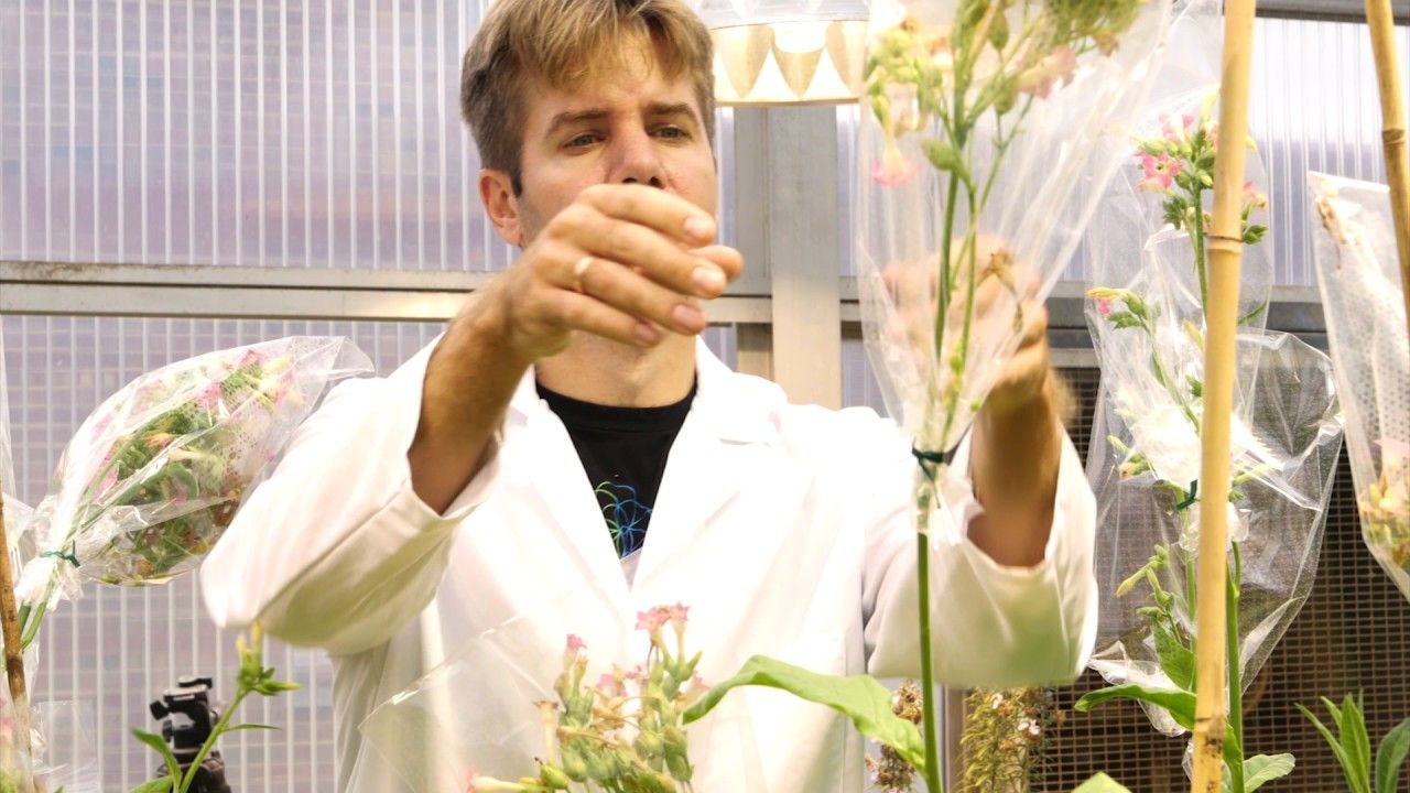
You could now 3D print your own tiny walking “bio-bots” powered by living muscle cells and controlled with electrical and light pulses, thanks to a new gennext robot ‘recipe’ developed by scientists.

This can result in exciting possibilities where these “systems could one day demonstrate complex behaviours including self-assembly, self-organisation, self-healing, and adaptation of composition and functionality to best suit their environment,” researchers said.

“The protocol teaches every step of building a bio-bot, from 3D printing the skeleton to tissue engineering the skeletal muscle actuator, including manufacturers and part numbers for every single thing we use in the lab,” said Ritu Raman, a postdoctoral fellow at the University of Illinois at Urbana-Champaign in the US.

Read more

By Julie Steenhuysen

CHICAGO (Reuters) — Powerful gene editing tools may one day be used on human embryos, eggs and sperm to remove genes that cause inherited diseases, according to a report by U.S. scientists and ethicists released on Tuesday.

The report from the National Academy of Sciences (NAS) and the National Academy of Medicine said scientific advances make gene editing in human reproductive cells “a realistic possibility that deserves serious consideration.”

Read more

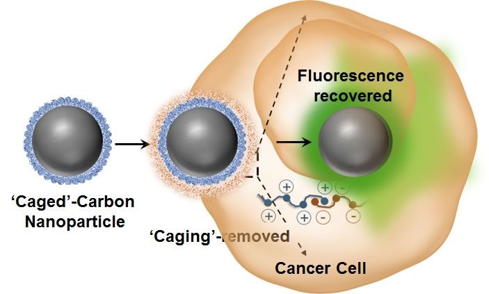
‘Caged’ non-fluorescent carbon dot enters the cancer cell, loses its caging and lights up. Credit: University of Illinois.

Tiny carbon dots have, for the first time, been applied to intracellular imaging and tracking of drug delivery involving various optical and vibrational spectroscopic-based techniques such as fluorescence, Raman, and hyperspectral imaging. Researchers from the University of Illinois at Urbana-Champaign have demonstrated, for the first time, that photo luminescent carbon nanoparticles can exhibit reversible switching of their optical properties in cancer cells.

“One of the major advantages of these agents are their strong intrinsic optical sensitivity without the need for any additional dye/fluorophore and with no photo-bleaching issues associated with it,” explained Dipanjan Pan, an assistant professor of bioengineering and the leader of the study. “Using some elegant nanoscale surface chemistry, we created a molecular ‘masking’ pathway to turn off the fluorescence and then selectively remove the mask leading to regaining the brightness.

Read more

A new story out on Engadget, emphasizing the need to make government treat science and technology as a primary focus:


Zoltan Istvan didn’t have much of a chance at being president, but that didn’t stop him from campaigning as the Transhumanist Party’s candidate to promote his pro-technology and science positions. Now, he’s setting his sights a bit lower, and with a different party. Istvan announced this morning that he plans to run for governor of California in 2018 under the Libertarian Party.

“We need leadership that is willing to use radical science, technology, and innovation—what California is famous for—to benefit us all,” he wrote in a Newsweek article. “We need someone with the nerve to risk the tremendous possibilities to save the environment through bioengineering, to end cancer by seeking a vaccine or a gene-editing solution for it, to embrace startups that will take California from the world’s 7th largest economy to maybe even the largest economy—bigger than the rest of America altogether.”

When we spoke to him in November, Istvan made it clear that he would be looking at the Libertarian Party if he were to run for president again. Not only does he identify as libertarian, he also saw the benefit of working with a more established political party, instead of starting one from the ground up.

“The most important thing I learned from my presidential campaign is that this is a team sport,” Istvan said in an email. “Without the proper managers, volunteers, spokespeople, and supporters, it’s really impossible to make a dent in an election. That’s part of the reason I joined the Libertarian Party for my governor run. They have tens of thousands of active supporters in California alone, so my election begins with real resources and infrastructure to draw upon. That’s a large difference from my Presidential campaign, where we essentially were shoe-stringing it the whole time.”

Read more

It looks like Self Driving cars may create a US organ shortage that finally acts as the Kick in the Ass to force stem cell generated organs on to the market. Enough of the ‘in the future’ we might have these Nonsesne.


Science, however, can offer better a better solution.

The waiting lists for donor organs are long — 120,000 people on a given day — and ever increasing. With fewer donor organs to go around, researchers are working on other ways to get people the parts they need. With help from 3D printing and other bioengineering technologies, we will eventually be able to grow our own organs and stop relying on donors.

Related: How Technology is Tackling Joe Biden’s Cancer Moonshot

“It’s like a race in which there’s multiple different players, but no matter which one of them winds up winning, it’s good,” says Ali Khademhosseini, a bioengineer at Harvard Medical School.

Read more

In Brief

  • Researchers were able to increase the weight of tobacco plants by around 14 to 20 percent compared to unmodified plants by adding in genes to improve the process of photosynthesis.
  • If successful with other plants, the method could improve the yields of food crops such as cowpeas, rice, and cassava and decrease world hunger.

In terms of plants, the concept of genetically modified organisms (GMOs) often refers to the insertion of genetic information from one species of plant to another so that the recipient plant gains a desirable trait. This process has been used extensively to improve crop yields. For example, one type of rice has been made waterproof so that yields aren’t affected by heavy floods from typhoons.

Now, a group of researchers from the University of California, Berkeley, has published a paper in the journal Science that takes a different approach to increasing crop yields. Instead of inserting genetic information from one species of plant into another to change it, these biologists used genetic modification to tweak a mechanism already inherent in plants.

Read more

Nice.


Testing treatments for bone cancer tumors may get easier with new enhancements to sophisticated support structures that mimic their biological environment, according to Rice University scientists.

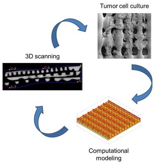
A team led by Rice bioengineer Antonios Mikos has enhanced its three-dimensional printed scaffold to see how Ewing’s sarcoma (bone cancer) cells respond to stimuli, especially shear stress, the force experienced by tumors as viscous fluid such as blood flows through bone. The researchers determined the structure of a scaffold, natural or not, has a very real effect on how cells express signaling proteins that help cancer grow.

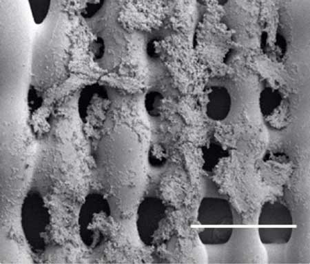
Sarcoma Cells on a scaffold

Sarcoma (bone cancer) cells proliferate on the surface of a 3D printed scaffold created at Rice University. Experiments at Rice showed that the size of pores in the scaffold, which mimics the extracellular matrix in bone, and the pores’ orientation make a difference in how cells proliferate in the presence of a flowing fluid, like blood. (Image: Mikos Research Group/Rice University)

Read more

This is definitely a share that is interesting to many studying synthetic organs and their acceptance into the human body as well as the work occurring on Quantum biology as well.


The goal of in vitro and in vivo toxicity testing is to identify compounds that would predict adverse reactions in humans. Olson et al. found that only 70% of human toxicity was predicted from animal testing. Currently we rely on traditional toxicity testing in animals, a 1930’s methodology that is now challenged due to questionable relevance to human risk, high cost, ethical concerns, and throughput that is too limited for the nearly 80,000 industrial chemicals not yet tested for safety. Additionally, testing usually extrapolates acute, high dose animal results to chronic, low dose human exposures, thereby risking rejection or limiting the use of drugs, industrial chemicals or consumer products. Moreover, the ability of lab animal target organ toxicity to predict dose-limiting toxicity in the corresponding human organ varies widely, from a low of 30% for human cutaneous toxicity, to 50–60% for human hepatotoxicity, to a high of 90% for hematological drug toxicity. Animal drug efficacy models are also notoriously discordant. In an analysis of six drugs to treat head injury, hemorrhage, acute ischemic stroke, neonatal respiratory distress syndrome, and osteoporosis, it was found that efficacy was similar in animals and humans for three drugs but was dissimilar for another three. In oncology drug development, animal models often over-predict anti-tumor efficacy in humans3,4. Examples such as these highlight the need to continue research into methods that reduce the dependence on laboratory animals for toxicity testing of environmental chemicals, determine efficacy and toxicity in drug development, serve as a mimic of human diseases, and provide patient-specific guidance in the emerging field of precision medicine.

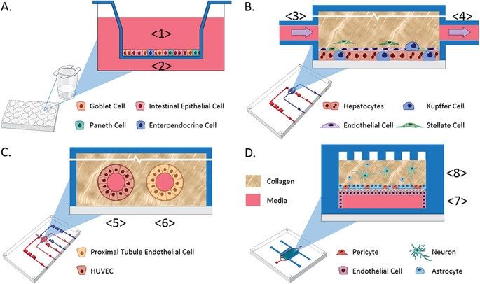
Recent advances in bioengineered materials, microfluidic technology, and the availability of human primary, immortalized, and induced pluripotent stem cell (iPSC)-derived cells are enabling development of human microphysiological systems (MPS), sometimes called “organs-on-a-chip” or “human-on-a-chip,” that use multiple organ-specific human cells to recapitulate many functional and structural properties of a human organ. It is now generally accepted and supported by data that cellular responses to drugs in most human organs are more accurately approximated in 3D cell cultures than in traditional static 2D cell cultures5,6. Microfluidic perfusion further improves model performance by providing a flow of nutrients and oxygen and the removal of waste products from the cell cultures. Physiologically relevant flow increases oxygen consumption, Krebs cycle activity and secretion of synthesized proteins, and decreases expression of the hypoxia HIF1 gene. Flow also improves the absorption and metabolism of compounds like benzo[a]pyrene6,8,9. The large number of recent publications reviewing organ MPS models indicates a high degree of interest by industrial and academic researchers, granting agencies and other stakeholders10,11,12,13. In addition to the stand-alone MPS, investigators are linking MPS to study organ-organ functional interactions, efficacy, PK and toxicology14,15,16,17,18.

An obvious approach to linking organs is direct coupling of the media stream outflow from one organ into the inflow of the next by use of tubing or a connecting channel. Some limitations to this approach include the requirement for a common medium, difficulty in reducing metabolic wastes to the next organ, organ-specific flow rates and adequate oxygenation of all modules in the system19. These requirements are most easily addressed when the linked organ modules are designed and developed at the same time and in the same laboratory, but even when the organ modules are co-developed, the proper scaling between organ modules is a significant design and calculation challenge. Although organ modules can be sized using allometric scaling20, the resulting functional capacity of the individual organ models may not scale the same.

Read more