Toggle light / dark theme

Researchers from Harvard University have 3D printed a school of soft robotic fish that are capable of swimming in complex patterns without the aid of Wi-Fi or GPS.

Inspired by the distinctive reef-dwelling surgeonfish, the team’s ‘Bluebots’ feature four fins for precision navigation, and a system of LEDs and cameras that enable them to swarm without colliding. The self-sufficiency of the tiny bots could make them ideal for ecological monitoring applications, in areas that wouldn’t otherwise be accessible to humans.

“Just by observing how far or close they are in a picture, they know how far or close the robot must be in the real world. That’s the trick we play here,” the study’s lead author Florian Berlinger told Wired.

Scientists from UNSW Sydney have developed a ceramic-based ink that may allow surgeons in the future to 3D-print bone parts complete with living cells that could be used to repair damaged bone tissue.

Using a 3D-printer that deploys a special ink made up of calcium phosphate, the scientists developed a new technique, known as ceramic omnidirectional bioprinting in cell-suspensions (COBICS), enabling them to print -like structures that harden in a matter of minutes when placed in water.

While the idea of 3D-printing bone-mimicking structures is not new, this is the first time such material can be created at room temperature—complete with living cells—and without harsh chemicals or radiation, says Dr. Iman Roohani from UNSW’s School of Chemistry.

Video. And it’s only the first step. Imagine a satellite that doesn’t need to rely on components it brought from earth. It can print out components for itself and for others; spare parts or upgrades for itself, other satellites and space stations.


Made In Space is building a satellite that can 3D print itself in space. If successful, their satellite could revolutionize how we design future spacecraft.

This may be good news for those who have damaged joints due to sports or old age.

😃


Human knees are notoriously vulnerable to injury or wearing out with age, often culminating in the need for surgery. Now researchers have created new hybrid bioinks that can be used to 3D print structures to replace damaged cartilage in the knee.

The meniscus is the rubbery cartilage that forms a C-shaped cushion in your knee, preventing the bones of your upper and lower leg from rubbing against each other. This stuff is susceptible to damage from sports injuries, but can also wear out with age – and if it gets particularly bad, sometimes the only thing left to do is surgically remove some of the damaged meniscus.

For the new proof-of-concept study, researchers at the Wake Forest Institute for Regenerative Medicine (WFIRM) demonstrated a new method for 3D bioprinting that creates both the cartilage and the supporting structures. The team used the Integrated Tissue and Organ Printing System (ITOPS), which has been used in past studies to print complex tissues such as bones, muscles and even ears.

Forget glue, screws, heat or other traditional bonding methods. A Cornell University-led collaboration has developed a 3D printing technique that creates cellular metallic materials by smashing together powder particles at supersonic speed.

This form of technology, known as “cold spray,” results in mechanically robust, that are 40% stronger than similar materials made with conventional manufacturing processes. The structures’ small size and porosity make them particularly well-suited for building biomedical components, like replacement joints.

The team’s paper, “Solid-State Additive Manufacturing of Porous Ti-6Al-4V by Supersonic Impact,” published Nov. 9 in Applied Materials Today.

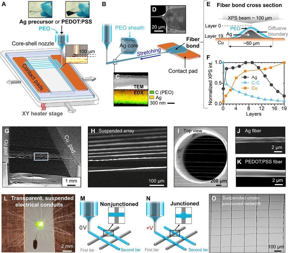
Scalability and device integration have been prevailing issues limiting our ability in harnessing the potential of small-diameter conducting fibers. We report inflight fiber printing (iFP), a one-step process that integrates conducting fiber production and fiber-to-circuit connection. Inorganic (silver) or organic {PEDOT: PSS [poly(3,4-ethylenedioxythiophene) polystyrene sulfonate]} fibers with 1- to 3-μm diameters are fabricated, with the fiber arrays exhibiting more than 95% transmittance (350 to 750 nm). The high surface area–to–volume ratio, permissiveness, and transparency of the fiber arrays were exploited to construct sensing and optoelectronic architectures. We show the PEDOT: PSS fibers as a cell-interfaced impedimetric sensor, a three-dimensional (3D) moisture flow sensor, and noncontact, wearable/portable respiratory sensors. The capability to design suspended fibers, networks of homo cross-junctions and hetero cross-junctions, and coupling iFP fibers with 3D-printed parts paves the way to additive manufacturing of fiber-based 3D devices with multilatitude functions and superior spatiotemporal resolution, beyond conventional film-based device architectures.

Small-diameter conducting fibers have unique morphological, mechanical, and optical properties such as high aspect ratio, low bending stiffness, directionality, and transparency that set them apart from other classes of conducting, film-based micro/nano structures (1–3). Orderly assembling of thin conducting fibers into an array or three-dimensional (3D) structures upscales their functional performance for device coupling. Developing new strategies to control rapid synthesis, patterning, and integration of these conducting elements into a device architecture could mark an important step in enabling new device functions and electronic designs (4, 5). To date, conducting micro/nanoscaled fibers have been produced and assembled in a number of ways, from transferring of chemically grown nanofibers/wires (6, 7), writing electrohydrodynamically deposited lines (8, 9), to drawing ultralong fibers (10, 11), wet spinning of fibers (12–14), and 2D/3D direct printing (15–18).

Researchers at the National Institute of Standards and Technology (NIST) have developed a new method of 3D-printing gels and other soft materials. Published in a new paper, it has the potential to create complex structures with nanometer-scale precision. Because many gels are compatible with living cells, the new method could jump-start the production of soft tiny medical devices such as drug delivery systems or flexible electrodes that can be inserted into the human body.

A standard 3D printer makes solid structures by creating sheets of material — typically plastic or rubber — and building them up layer by layer, like a lasagna, until the entire object is created.

Using a 3D printer to fabricate an object made of gel is a “bit more of a delicate cooking process,” said NIST researcher Andrei Kolmakov. In the standard method, the 3D printer chamber is filled with a soup of long-chain polymers — long groups of molecules bonded together — dissolved in water. Then “spices” are added — special molecules that are sensitive to light. When light from the 3D printer activates those special molecules, they stitch together the chains of polymers so that they form a fluffy weblike structure. This scaffolding, still surrounded by liquid water, is the gel.