Now just need to go to rat monkey human.
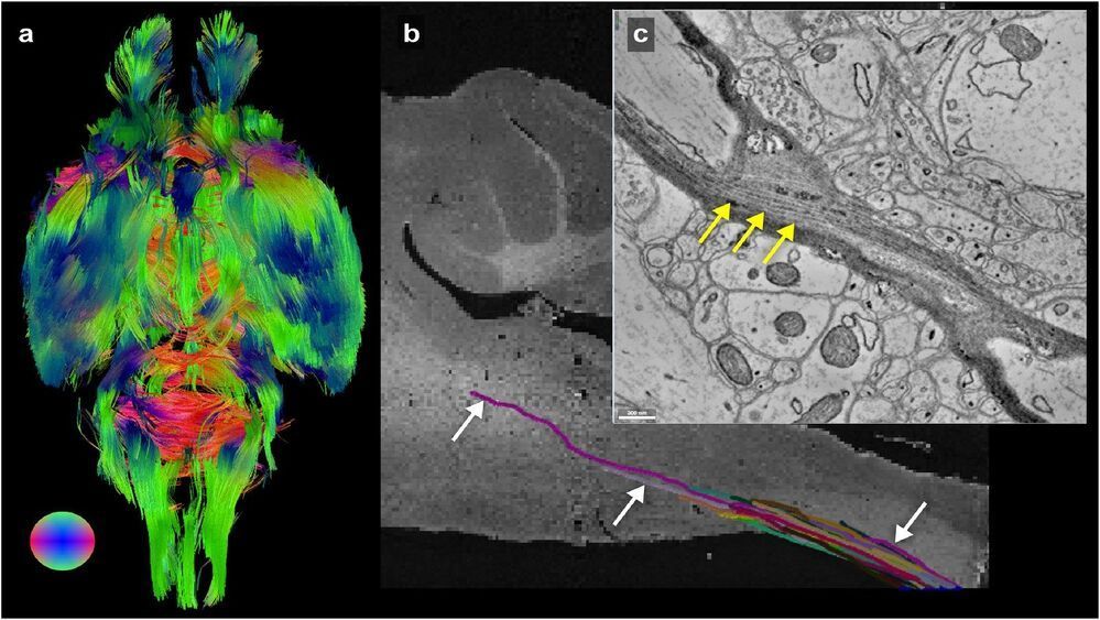
Researchers at the University of Chicago and the U.S. Department of Energy’s (DOE) Argonne National Laboratory have imaged an entire mouse brain across five orders of magnitude of resolution, a step which researchers say will better connect existing imaging approaches and uncover new details about the structure of the brain.
The advance, which was published on June 9 in NeuroImage, will allow scientists to connect biomarkers at the microscopic and macroscopic level. It leveraged existing advanced X-ray microscopy techniques at the Advanced Photon Source (APS), a DOE Office of Science User Facility at Argonne, to bridge the gap between MRI and electron microscopy imaging, providing a viable pipeline for multiscale whole brain imaging within the same brain.
“Argonne had this extremely powerful X-ray microscope, and it hadn’t been used for brain mapping yet, so we decided to try it out.” — Assistant Professor Bobby Kasthuri
