O,.,o.
Since their invention in the 1930s, electron microscopes have helped scientists peer into the atomic structure of ordinary materials like steel, and even exotic graphene. But despite these advances, such imaging techniques cannot precisely map out the 3D atomic structure of materials in a liquid solution, such as a catalyst in a hydrogen fuel cell, or the electrolytes in your car’s battery.
Now, researchers at Berkeley Lab, in collaboration with the Institute for Basic Science in South Korea, Monash University in Australia, and UC Berkeley, have developed a technique that produces atomic-scale 3D images of nanoparticles tumbling in liquid between sheets of graphene, the thinnest material possible.
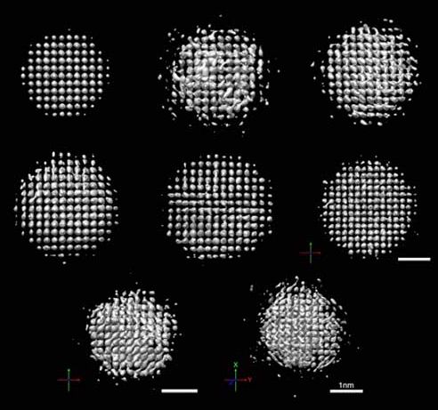
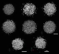
3D images of platinum particles between 2–3 nm in diameter shown rotating in liquid under an electron microscope. Each nanoparticle has approximately 600 atoms. White spheres indicate the position of each atom in a nanoparticle. (Image: IBS)
Amazing