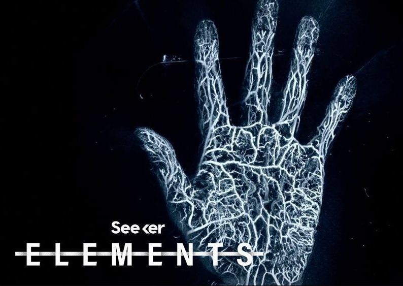
While X-rays can produce harmful radiation, a new technique using laser-induced sound waves provides highly detailed images of the structures in our bodies.
» Subscribe to Seeker!http://bit.ly/subscribeseeker
» Watch more Elements! http://bit.ly/ElementsPlaylist
Photoacoustic imaging is an emerging imaging technique that shoots micro-pulses of laser light at a specimen or body part, which selectively heats up parts of the tissue causing them to expand, and generate waves of pressure – a.k.a. sound waves.
Ultrasonic sensors are situated to capture these microscopic changes, and a processing software then reconstructs the image based on what the sensors “hear.” The speed of the laser can be adjusted depending on what type of tissue one would like to visualize.
The photoacoustic imaging technique is beginning to take off in both the medical and scientific worlds, as it provides us with super clear, incredibly detailed images of the human body and the structures inside it.
Not to mention, the imaging technique causes no discomfort and there is no dangerous ionizing radiation involved, making it a desirable alternative to more traditional imaging, like a CT scan, ultrasound, or a PET scan.
Not only can this new imaging technology be used to image tissues at extremely high resolution, you can also introduce a foreign material, like a contrast dye or a specially designed nanoparticle, to see things you might not be able to otherwise.
