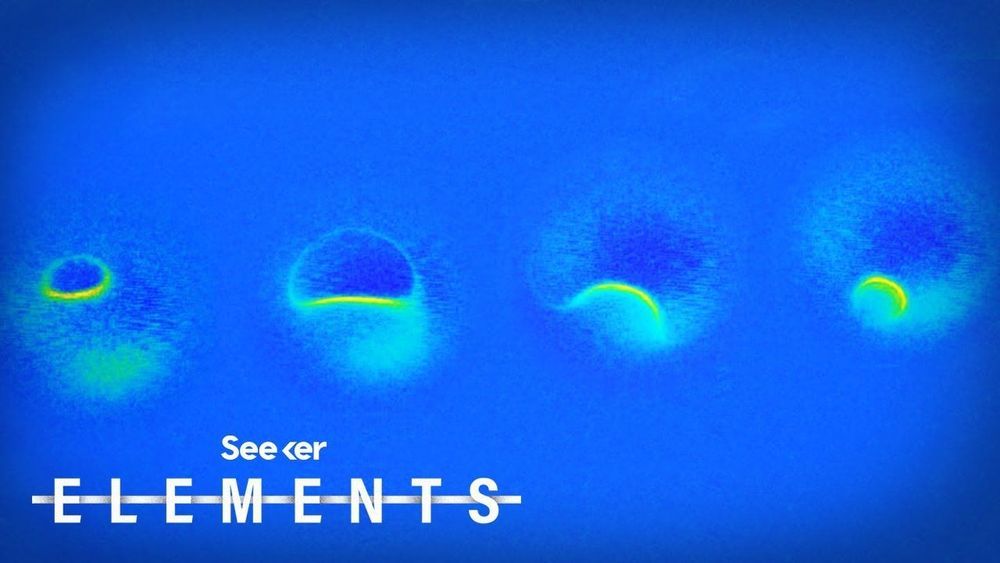
Magnetic resonance imaging is nothing new, but scientists were able to perform an MRI on a single atom. But how?
» Subscribe to Seeker! http://bit.ly/subscribeseeker
» Watch more Elements! http://bit.ly/ElementsPlaylist
Scientists recently captured the smallest MRI ever while scanning an individual atom. The technique successfully reached a breakthrough level of resolution in the world of microscopy, the detailed MRI can reveal single atoms as well as different types of atoms based on their magnetic interactions.
This breakthrough has potential applications in all kinds of fields, like quantum computing where it could be used to design atomic-scale methods of storing info or when it comes to drug development, the ability to control individual atoms could potentially be used to study how proteins fold and then lead to the development of drugs for diseases like Alzheimers.
In a sense, the researchers combined a version of an MRI machine with a special instrument called a scanning tunneling microscope, which turned out to be a match made in microscopy heaven.
An MRI scanner creates an extremely strong magnetic field around whatever it’s trying to image, temporarily re-aligning the protons in your body with that magnetic field. Then the MRI machine pulses the sample (or patient) with a radiofrequency, which pulls the protons slightly out of alignment with the magnetic field. And after the brief radiofrequency pulse is over, the protons snap back into alignment with the field, and the energy that’s released as the protons move back into place with the magnetic field is what is detected and visualized by the machine.
And a scanning tunneling microscope is used for imaging really tiny surfaces, and it can pick up certain properties like size and molecular structure.
