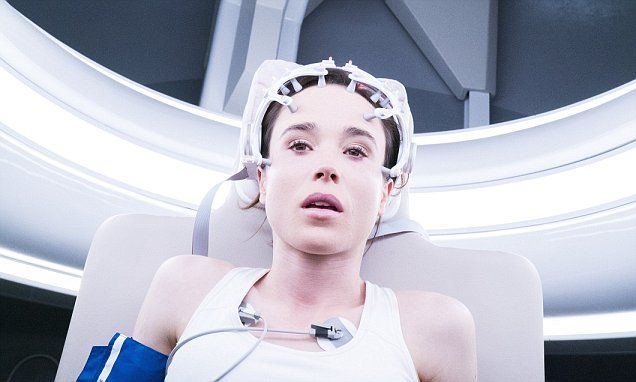
Alzheimer’s may be contagious.
A protein might be capable of spreading Alzheimer’s through blood transfusions and surgical equipment, but we don’t know yet how much of a risk this is.
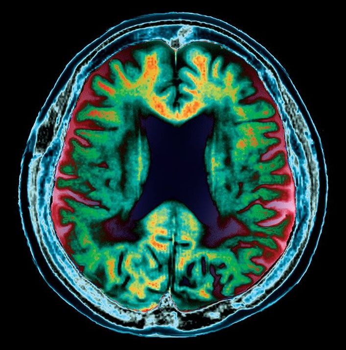
PROFITABLY recycling waste is always a good idea. And the Allen Institute for Brain Science, in Seattle, has found a way to recycle what is perhaps the most valuable waste of all—living human brain tissue. Understandably, few people are willing to donate parts of their brains to science while they are still alive. But, by collaborating with seven local neurosurgeons, the institute’s chief scientist, Christof Koch, and his colleagues, have managed to round up specimens of healthy tissue removed by those surgeons in order to get to unhealthy parts beyond them, which needed surgical ministration. Normally, such tissue would be disposed of as waste. Instead, Dr Koch is making good use of it.
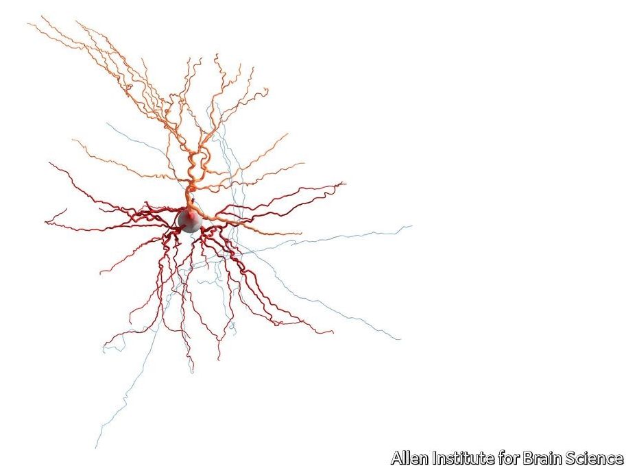
The repository the cells from these samples end up in is a part of a wider project, the Allen Cell Types Database. The first data from the newly collected human brain cells were released on October 25th. The Allen database, which is open for anyone to search, thus now includes information on the shape, electrical activity and gene activity of individual human neurons. The electrical data are from 300 live neurons of various types, taken from 36 people. The shapes (see picture for example) are from 100 of these neurons. The gene-expression data come from 16,000 neurons, though those cells are post-mortem samples.
The human brain is the most complex object in the known universe. Because it is more complicated than animal brains in ways that (say) human livers are not more complicated than animal livers, using animal brains as analogues of human ones is never going to be satisfactory. Dr Koch’s new database may therefore help explain what is special about human brains. That will assist understanding of brain diseases and disorders. It may also shed light on one of his particular interests, the nature of consciousness.
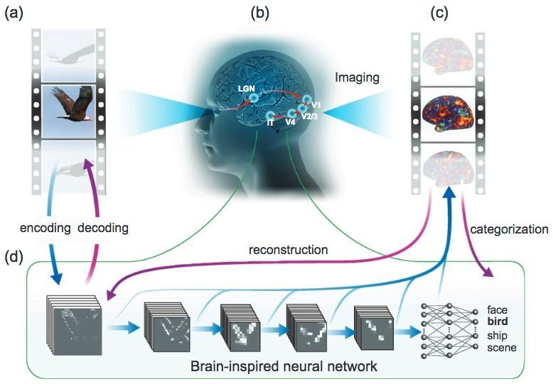
Purdue Engineering researchers have developed a system that can show what people are seeing in real-world videos, decoded from their fMRI brain scans — an advanced new form of “mind-reading” technology that could lead to new insights in brain function and to advanced AI systems.
The research builds on previous pioneering research at UC Berkeley’s Gallant Lab, which created a computer program in 2011 that translated fMRI brain-wave patterns into images that loosely mirrored a series of images being viewed.

The relationship between the mind and the brain is a mystery that is central to how we understand our very existence as sentient beings. Some say the mind is strictly a function of the brain — consciousness is the product of firing neurons. But some strive to scientifically understand the existence of a mind independent of, or at least to some degree separate from, the brain.
The peer-reviewed scientific journal NeuroQuantology brings together neuroscience and quantum physics — an interface that some scientists have used to explore this fundamental relationship between mind and brain.
An article published in the September 2017 edition of NeuroQuantology reviews and expands upon the current theories of consciousness that arise from this meeting of neuroscience and quantum physics.

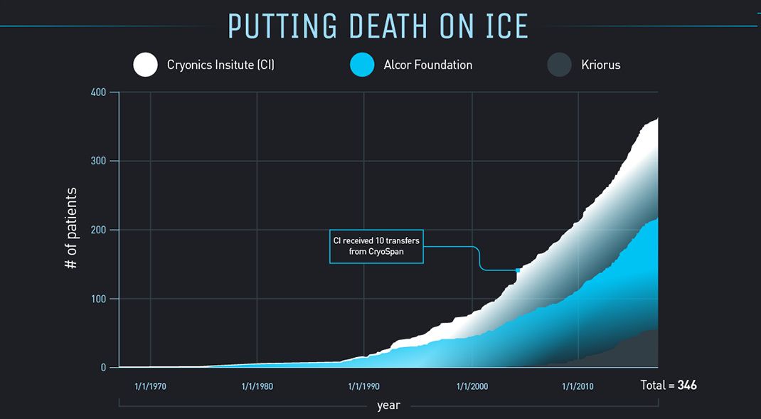
Robert C. W. Ettinger’s seminal work, The Prospect Of Immortality, detailed many of the scientific, moral, and economic implications of cryogenically freezing humans for later reanimation. It was after that book was published in 1962 that the idea of freezing one’s body after death began to take hold.
One of the most pressing questions is, even if we’re able to revive a person who has been cryogenically preserved, will the person’s memories and personality remain intact? Ettinger posits that long-term memory is stored in the brain as a long-lasting structural modification. Basically, those memories will remain, even if the brain’s “power is turned off”.
This infographic delves into the mechanics and feasibility of cryonics – a process that thousands of people are betting will give them a second shot at life.
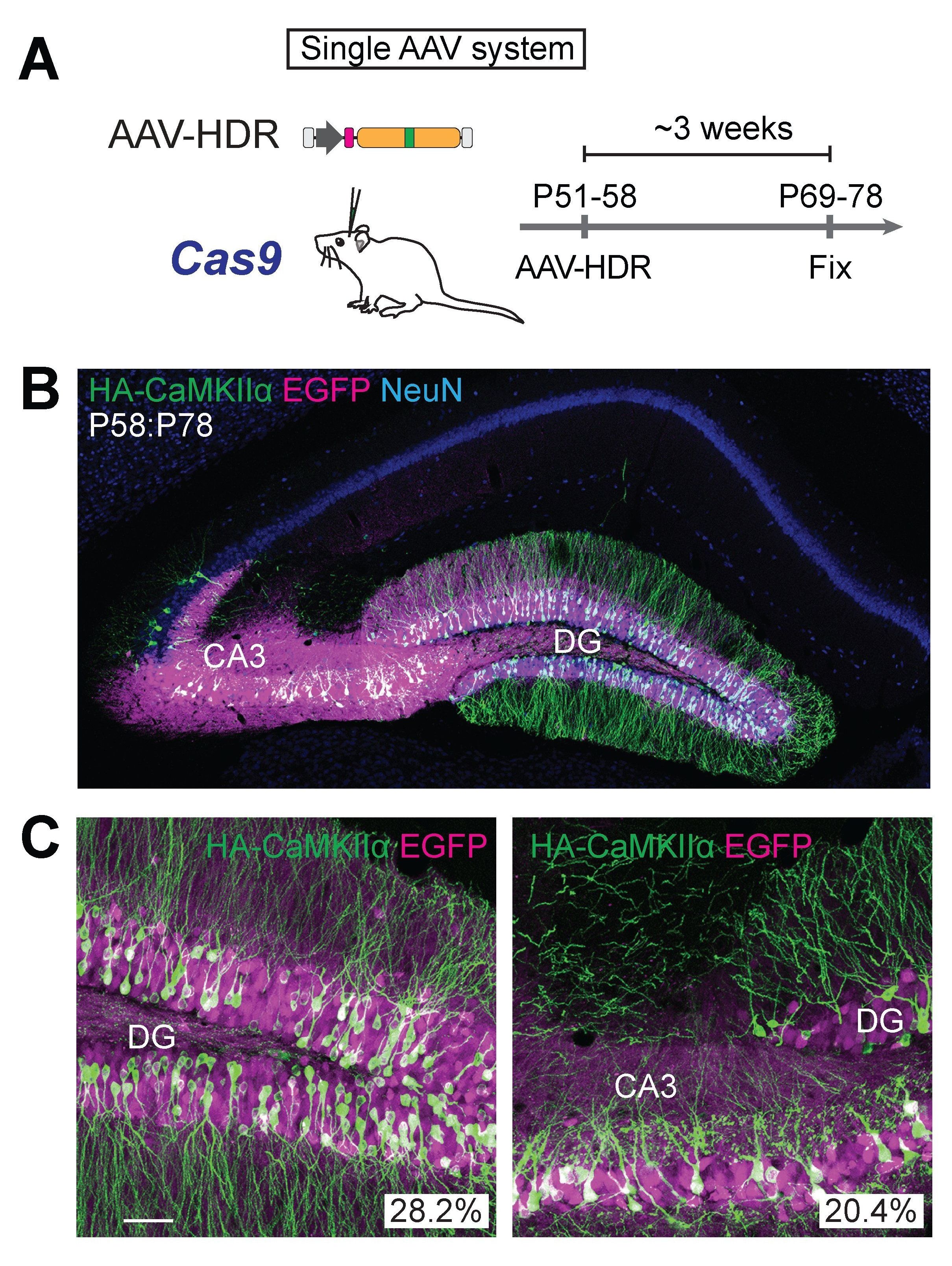
Genome editing technologies have revolutionized biomedical science, providing a fast and easy way to modify genes. However, the technique allowing scientists to carryout the most precise edits, doesn’t work in cells that are no longer dividing — which includes most neurons in the brain. This technology had limited use in brain research, until now. Research Fellow Jun Nishiyama, M.D., Ph.D., Research Scientist, Takayasu Mikuni, M.D., Ph.D., and Scientific Director, Ryohei Yasuda, Ph.D. at the Max Planck Florida Institute for Neuroscience (MPFI) have developed a new tool that, for the first time, allows precise genome editing in mature neurons, opening up vast new possibilities in neuroscience research.
This novel and powerful tool utilizes the newly discovered gene editing technology of CRISPR-Cas9, a viral defense mechanism originally found in bacteria. When placed inside a cell such as a neuron, the CRISPR-Cas9 system acts to damage DNA in a specifically targeted place. The cell then subsequently repairs this damage using predominantly two opposing methods; one being non-homologous end joining (NHEJ), which tends to be error prone, and homology directed repair (HDR), which is very precise and capable of undergoing specified gene insertions. HDR is the more desired method, allowing researchers flexibility to add, modify, or delete genes depending on the intended purpose.
Coaxing cells in the brain to preferentially make use of the HDR DNA repair mechanism has been rather challenging. HDR was originally thought to only be available as a repair route for actively proliferating cells in the body. When precursor brain cells mature into neurons, they are referred to as post-mitotic or nondividing cells, making the mature brain largely inaccessible to HDR — or so researchers previously thought. The team has now shown that it is possible for post-mitotic neurons of the brain to actively undergo HDR, terming the strategy “vSLENDR (viral mediated single-cell labeling of endogenous proteins by CRISPR-Cas9-mediated homology-directed repair).” The critical key to the success of this process is the combined use of CRISPR-Cas9 and a virus.
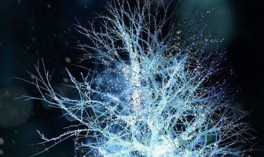
Researchers have developed a technique that enables gene editing on neurons — something previously thought to be impossible. This new tool will present amazing new opportunities for neuroscience research.
Technologies designed for editing the human genome are transforming biomedical science and providing us with relatively simple ways to modify and edit genes. However, precision editing has not been possible for cells that have stopped dividing, including mature neurons. This has meant that gene editing has been of limited use in neurological research — until now. Researchers at the Max Planck Florida Institute for Neuroscience (MPFI) have created a new tool that allows, for the first time ever, precise genome editing in mature neurons. This relieves previous constraints and presents amazing new opportunities for neuroscience research.
