Toggle light / dark theme

Very promising since “Identifying what changes are happening in the brain when interventions successfully reduce depressive symptoms could allow us to create more effective, pharmaceutical-free approaches to help alleviate depression in people who experience chronic traumatic brain injury symptoms,” said study author Dr. Sandra Bond Chapman, founder and chief director of the Center for BrainHealth.


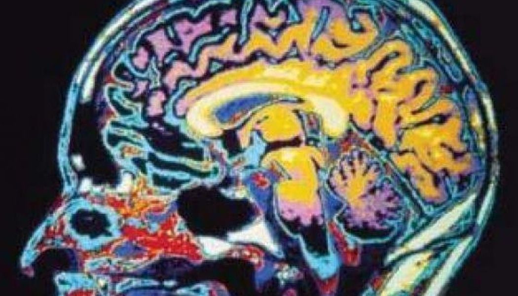
Images show prefrontal connectivity patterns after cognitive training in individuals who suffered traumatic brain injury. Kihwan Han et al (2018) _____ Cognitive training reduces depression, rebuilds injured brain structure & connectivity after traumatic brain injury (UT-Dallas release): “New research from the Center.

Read more

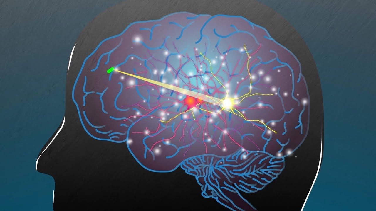
MIT researchers, working with scientists from Brigham and Women’s Hospital, have developed a new way to power and communicate with devices implanted deep within the human body. Such devices could be used to deliver drugs, monitor conditions inside the body, or treat disease by stimulating the brain with electricity or light.

The implants are powered by radio frequency waves, which can safely pass through human tissues. In tests in animals, the researchers showed that the waves can power devices located 10 centimeters deep in tissue, from a distance of 1 meter.

“Even though these tiny implantable devices have no batteries, we can now communicate with them from a distance outside the body. This opens up entirely new types of medical applications,” says Fadel Adib, an assistant professor in MIT’s Media Lab and a senior author of the paper, which will be presented at the Association for Computing Machinery Special Interest Group on Data Communication (SIGCOMM) conference in August.

Read more

New research on brain ageing and mitochondria from Salk Institute.


Thanks to a new technique, researchers from the Salk Institute’s Gage laboratory have shown that impaired energy production might be a reason why human brains are susceptible to age-related diseases in the first place [1].

In particular, Salk scientists observed that induced neurons (iNs) obtained from fibroblasts of older individuals had dysfunctional mitochondria and therefore decreased energy levels compared to younger neurons. Out-of-shape mitochondria have previously been implicated in degenerative brain diseases, such as Alzheimer’s and Parkinson’s, and this finding might help reveal more about the connection between these diseases and this particular hallmark of aging.

Mitochondrial dysfunction 101

Our readers are probably familiar with the 2013 study “The Hallmarks of Aging”, a review describing in detail what is known of the typical signs of age-related degeneration at the molecular level [2]. Mitochondrial dysfunction is a hallmark in its own right, and it can be thought of as the meltdown of cellular energy production facilities.

Read more

http://www.thepathmag.com/can-you-actually-hack-your-dna-to-slow-down-aging/

Many technologies / interventions progressing down the development pathways in the coming years — but there are a lot of free, common sense adjustments you can make today:

http://www.blogtalkradio.com/entrepreneursempower/2018/06/03/free-your-mind-with-trease-smith-guest-ira-pastor

I t’s an odd thing for someone to say about neurons: “Let’s see if anyone is awake.” And it’s an even odder thing to hear in a cavernous, half-furnished office suite where one whole room is occupied only by copy machines and a lonely foosball table.

Not far from that foosball table, Oshiorenoya Agabi and Benjamin Sadrian are sitting in a lab at their startup, Koniku, in Berkeley, California. Agabi founded the company, and Sadrian is a senior neuroscientist. They are toggling between a microscope and a screen full of blue graphs, looking for signs of activity in a cluster of neurons. Sadrian pauses as he scrolls through slightly fuzzy readouts on the screen, reminiscent of stock charts with buzz cuts. “I wish you’d come later, even tomorrow,” he sighs.

These readouts measure signals inside cells, and Agabi and Sadrian are looking for spikes that would show Koniku’s neurons reacting to a chemical Sadrian exposed them to moments ago. When we examined them under the microscope, they glowed a faint neon green, which indicates they’re starting to mature. A few tentative dendrites reached out into the void, the neurons just beginning to form connections with one another. But the telltale spikes don’t materialize on the screen. At just six days old, these neurons are still too young to do the jobs they’ve been engineered to do.

Read more

With the ability to be coaxed into different kinds of mature cell types, induced pluripotent stem cells (iPSCs) hold all kinds of potential in the world of regenerative medicine. One of the many possibilities could be repairing damaged hearts, something that will soon be put to the test for the first time ever in newly approved clinical trials in Japan.

Since emerging from the laboratory of researcher Shinya Yamanaka in Japan in 2006, the potential of iPSCs has been explored in all kinds of promising research efforts. We have seen them implanted into rabbits to restore their vision, become brain tumor predators, and turned into precursor cells for human organs.

IPSCs are created by first harvesting cells from body tissues and then infecting them with a virus, in turn introducing them to carefully selected genes that return them to their immature state. From there they can develop into any cell in the body, a capability so powerful it earned Yamanaka a Nobel Prize in 2012.

Read more

On Thursday, an international team of scientists announced a new explanation for the evolution of brain size expansion: Unique genes that, to date, have only been found in humans. They’re called NOTCHN2NL genes and so far the scientists have identified eight different versions of them.

Read more

https://www.engadget.com/…/3D-printed-brain-medical-imagin…/


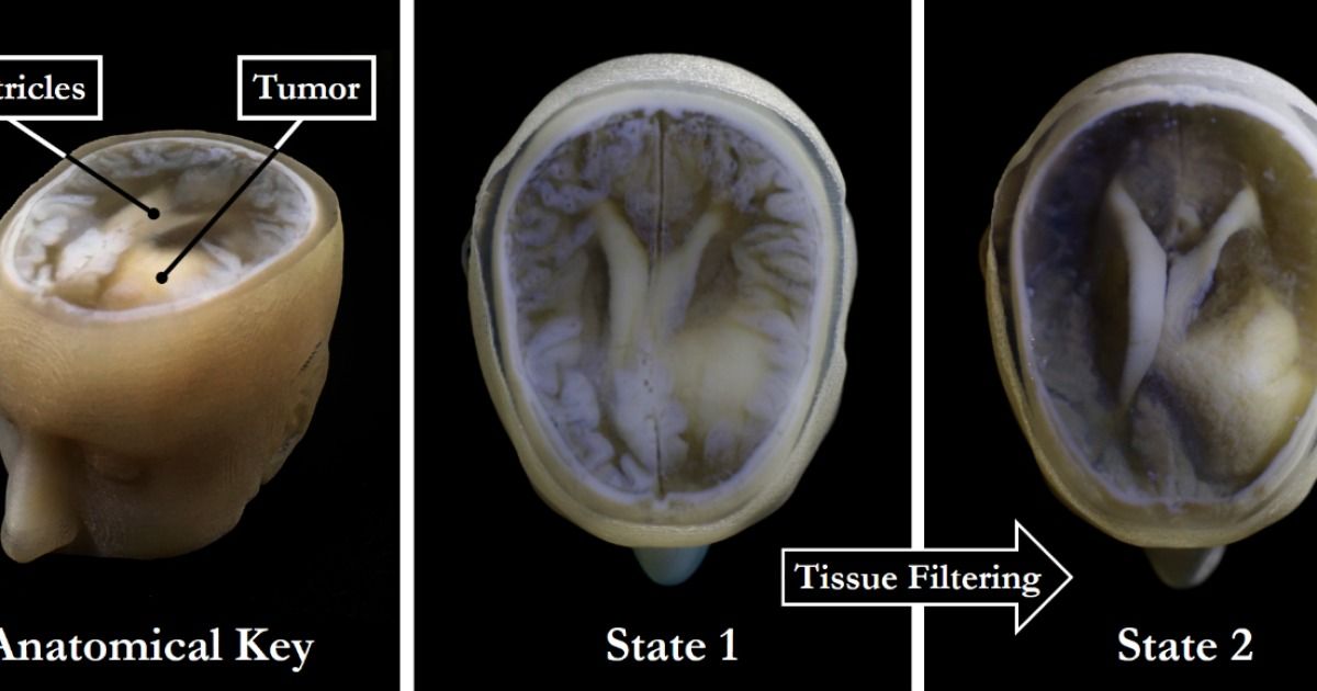
There are almost limitless possibilities when it comes to 3D printing. Design your own color-changing jewelry? Fine. Fabricate your own drugs? No problem. Print an entire house in under 24 hours? Sure! Now, researchers have come up with a fast and easy way to print palm-sized models of individual human brains, presumably in a bid to advance scientific endeavours, but also because, well, that’s pretty neat.

In theory, creating a 3D printout of a human brain has been done before, using data from MRI and CT scans. But as MIT graduate Steven Keating found when he wanted to examine his own brain following his surgery to remove a baseball-sized tumour, it’s a slow, cumbersome process that doesn’t reveal any important areas of interest.

MRI and CT scans produce images with so much detail that objects of interest need to be isolated from surrounding tissue and converted into surface meshes in order to be printed. This involves a radiologist manually tracing the desired object onto every single image “slice” of the brain, or it can be done via automatic thresholding, where a computer converts areas that contain grayscale pixels into either solid black or solid white pixels, based on a shade of gray that is chosen to be the threshold between black and white. But since medical imaging data often contains irregularly-shaped objects and lacks clear borders, features of interest are usually over- or under-exaggerated, and details are washed out.

Read more