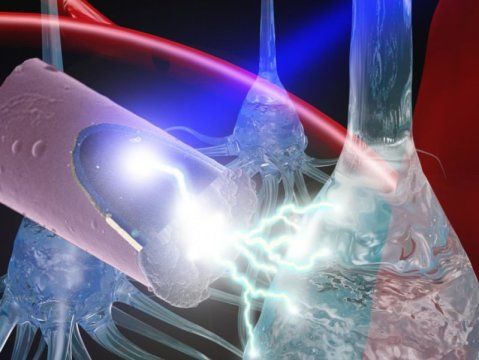
Neural stimulation is a developing technology that has beneficial therapeutic effects in neurological disorders, such as Parkinson’s disease. While many advancements have been made, the implanted devices deteriorate over time and cause scarring in neural tissue. In a recently published paper, the University of Pittsburgh’s Takashi D. Y. Kozai detailed a less invasive method of stimulation that would use an untethered ultrasmall electrode activated by light, a technique that may mitigate damage done by current methods.
Category: neuroscience
LA JOLLA—(February 18, 2019) Aging is a leading risk factor for a number of debilitating conditions, including heart disease, cancer and Alzheimer’s disease, to name a few. This makes the need for anti-aging therapies all the more urgent. Now, Salk Institute researchers have developed a new gene therapy to help decelerate the aging process.
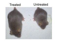
The findings, published on February 18, 2019 in the journal Nature Medicine, highlight a novel CRISPR/Cas9 genome-editing therapy that can suppress the accelerated aging observed in mice with Hutchinson-Gilford progeria syndrome, a rare genetic disorder that also afflicts humans. This treatment provides important insight into the molecular pathways involved in accelerated aging, as well as how to reduce toxic proteins via gene therapy.
“Aging is a complex process in which cells start to lose their functionality, so it is critical for us to find effective ways to study the molecular drivers of aging,” says Juan Carlos Izpisua Belmonte, a professor in Salk’s Gene Expression Laboratory and senior author of the paper. “Progeria is an ideal aging model because it allows us to devise an intervention, refine it and test it again quickly.”
And you my good sir the psychiatrist… according to the newer than new 6th edition of the Diagnostic and Statistical Manual…you have a labeling disorder. 🤓😋.
After nearly a century of effort, psychiatry’s best diagnoses leave much to be desired.
- By Daniel S. Barron on February 19, 2019
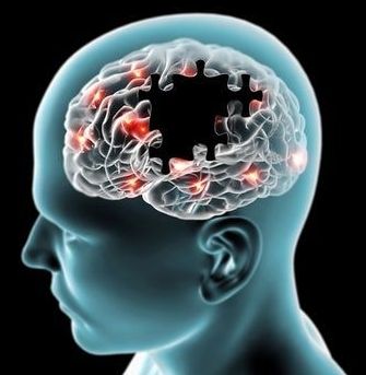
Daydreaming, thinking about the past, planning for the future, or just letting your mind wander off from the current moment occupies our thoughts for a large part of every day. However, a new study has revealed patients suffering from a specific kind of early-onset dementia may have completely lost the ability to do this and seem perpetually “stuck in the moment”.
Researchers have shown that a type of magnetic resonance imaging—called neuromelanin-sensitive MRI (NM-MRI)—is a potential biomarker for psychosis. NM-MRI signal was found to be a marker of dopamine function in people with schizophrenia and an indicator of the severity of psychotic symptoms in people with this mental illness. The study, funded by the National Institute of Mental Health (NIMH), part of the National Institutes of Health, appears in the Proceedings of the National Academy of Science.
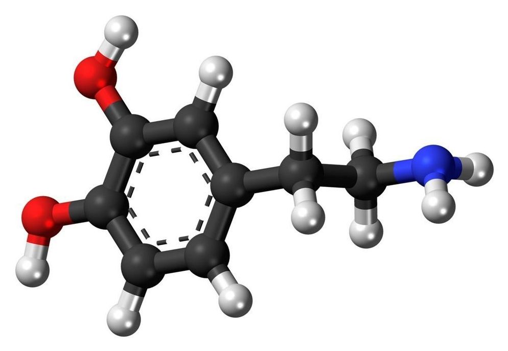
“Disturbances affecting the neurotransmitter dopamine are associated with a host of mental and neurological disorders, such as schizophrenia and Parkinson’s disease,” said Joshua A. Gordon, M.D., Ph.D., director of NIMH. “Because of the role dopamine plays in these disorders, the ability to measure dopamine activity is critical for furthering our understanding of these disorders, including how to best diagnose and treat them.”
Neuromelanin is a dark pigment created within dopamine neurons of the midbrain—particularly in the substantia nigra, a brain area that plays a role in reward and movement. Neuromelanin accumulates over the lifespan and is only cleared away from cells following cell death, as occurs in neurodegenerative disorders such as Parkinson’s disease. Researchers have found that NM-MRI signal is lower in the substantia nigra of people with Parkinson’s disease, reflecting the cell death that occurs in these patients.
Summary: Researchers have identified a previously unknown form of neural communication. They report the findings could help better the understanding of neural activity associated with specific brain processing and neurological disorders.
Source: Case Western Reserve University.
Biomedical engineering researchers at Case Western Reserve University say they have identified a previously unidentified form of neural communication, a discovery that could help scientists better understand neural activity surrounding specific brain processes and brain disorders.
Circa 2015
The first successful vaccination of deer against chronic wasting disease is reported in the journal Vaccine, (Vaccine 2015;38:726–33), posted online in advance of print Dec. 21, 2014.
Researchers say the breakthrough may not only protect U.S. livestock against CWD but may also shed new light on human diseases suspected of being caused by prion infections, such as Creutzfeldt-Jakob disease, kuru, familial insomnia, and variably protease-sensitive prionopathy. Some studies also have associated prionlike infections with Alzheimer’s disease.
“Now that we have found that preventing prion infection is possible in animals, it’s likely feasible in humans as well,” said senior study investigator and neurologist Thomas Wisniewski, MD, a professor at New York University’s Langone Medical Center.
It is the first such research to be undertaken at the university.
IBRO is the global federation of neuroscience organizations that aims to promote and support neuroscience around the world through training, teaching, collaborative research, outreach and advocacy.
The research will be carried out at Sahu’s Cell Biology and Molecular metabolism lab at the Vikram Sarabhai Institute of Cell and Molecular Biology, which is headed by Professor Sarita Gupta.
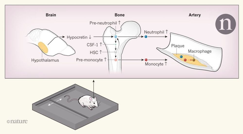
Most people have at some point echoed Macbeth’s complaint about the loss of “sleep that knits up the ravelled sleeve of care”. Sleep disorders, such as obstructive sleep apnoea (when breathing temporarily stops, causing both sleep disruption and lack of oxygen in blood) and sleep deprivation, have been associated with an increased risk of atherosclerosis and its harmful cardiovascular effects,. Atherosclerosis is characterized by the formation of ‘plaques’ in arteries, as white blood cells enter the artery wall, take up cholesterol and other substances from the blood and trigger an inflammatory response. However, the mechanisms linking sleep disruption and atherosclerosis have been largely unknown. Writing in Nature, McAlpine et al. show that persistent sleep disruption causes the brain to signal the bone marrow to increase the production of white blood cells.
McAlpine et al. studied mice that were prone to developing atherosclerosis. The authors induced sleep fragmentation by moving a bar intermittently across the bottom of the animals’ cages during their sleep period (Fig. 1), and compared these animals with animals that slept normally. They found that mice with sleep fragmentation had more-severe atherosclerosis, which was paralleled by increases in the production of white blood cells in the bone marrow and in the numbers of monocytes and neutrophils — two types of white blood cell — in the blood. These effects were absent if the bar was moved when the mice were fully awake. Stress activates the sympathetic nervous system (which is associated with the ‘fight-or-flight’ response), and such activation increases the production of white blood cells and atherosclerosis in other experimental settings. However, the authors did not find evidence for a role of sympathetic activation in their setting.