The mind is a powerful thing – it can generate both symptoms of illness and symptoms of healing. Here’s what this could tell us about consciousness.

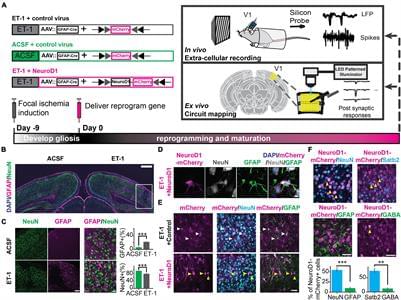
Neural circuits underlying brain functions are vulnerable to damage, including ischemic injury, leading to neuronal loss and gliosis. Recent technology of direct conversion of endogenous astrocytes into neurons in situ can simultaneously replenish the neuronal population and reverse the glial scar. However, whether these newly reprogrammed neurons undergo normal development, integrate into the existing neuronal circuit, and acquire functional properties specific for this circuit is not known. We investigated the effect of NeuroD1-mediated in vivo direct reprogramming on visual cortical circuit integration and functional recovery in a mouse model of ischemic injury. After performing electrophysiological extracellular recordings and two-photon calcium imaging of reprogrammed cells in vivo and mapping the synaptic connections formed onto these cells ex vivo, we discovered that NeuroD1 reprogrammed neurons were integrated into the cortical microcircuit and acquired direct visual responses. Furthermore, following visual experience, the reprogrammed neurons demonstrated maturation of orientation selectivity and functional connectivity. Our results show that NeuroD1-reprogrammed neurons can successfully develop and integrate into the visual cortical circuit leading to vision recovery after ischemic injury.
Functional circuit impairment associated with neuronal loss is commonly seen in patients with brain injuries, such as ischemia. Though neural stem cells (NSCs) exist in the subventricular zone (SVZ) in the adult brain, they are found to differentiate mainly into astrocytes when they migrate to injured cortex (Benner et al., 2013; Faiz et al., 2015), and their neurogenesis capacity is too limited to compensate for the neuronal loss. Currently, it still remains a challenge to generate neurons in adults and functionally incorporate them into the local circuits. Several strategies have shown the capability to induce neurogenesis and lead to some behavioral recovery. One promising approach is to transplant stem cell-derived neurons or neural progenitor cells (Tornero et al., 2013; Michelsen et al., 2015; Falkner et al., 2016; Somaa et al., 2017). Yet, there are concerns about graft rejection and tumorigenicity of the transplanted cells (Erdo et al., 2003; Marei et al., 2018).

Live human brain tissue — generously donated by brain surgery patients with epilepsy or tumors — is yielding incredible #neuroscience insights. A study on cells… See More.
As part of an international effort to map cell types in the brain, scientists identified increased diversity of neurons in regions of the human brain that expanded during our evolution.
Effort to scan the entire Human Brain continues.
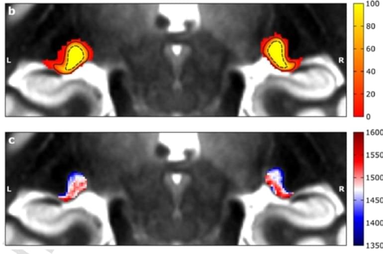
Summary: A new, non-invasive neuroimaging technique allowed researchers to investigate the visual sensory thalamus, a brain area associated with visual difficulties in dyslexia and other disorders.
Source: TU Dresden
The visual sensory thalamus is a key region that connects the eyes with the cerebral cortex. It contains two major compartments. Symptoms of many diseases are associated with alterations in this region. So far, it has been very difficult to assess these two compartments in living humans, because they are tiny and located very deep inside the brain.
This difficulty of investigating the visual sensory thalamus in detail has hampered the understanding of the function of visual sensory processing tremendously in the past. By coincidence, Christa Müller-Axt, Ph.D. student in the lab of neuroscientist Prof. Katharina von Kriegstein at TU Dresden, discovered structures that she thought might resemble the two visual sensory thalamus compartments in neuroimaging data.

“The use of organophosphate esters in everything from TVs to car seats has proliferated under the false assumption that they’re safe,” said Heather Patisaul, lead author and neuroendocrinologist at North Carolina State University. “Unfortunately, these chemicals appear to be just as harmful as the chemicals they’re intended to replace but act by a different mechanism.”
Summary: Exposure to even low levels of common chemicals called organophosphate esters can harm IQ, memory, learning, and brain development overall in young children.
Source: Green Science Policy Institute
Chemicals increasingly used as flame retardants and plasticizers pose a larger risk to children’s brain development than previously thought, according to a commentary published today in Environmental Health Perspectives.
The research team reviewed dozens of human, animal, and cell-based studies and concluded that exposure to even low levels of the chemicals—called organophosphate esters—may harm IQ, attention, and memory in children in ways not yet looked at by regulators.

It probably didn’t feel like much, but that simple kind of motion required the concerted effort of millions of different neurons in several regions of your brain, followed by signals sent at 200 mph from your brain to your spinal cord and then to the muscles that contracted to move your arm.
At the cellular level, that quick motion is a highly complicated process and, like most things that involve the human brain, scientists don’t fully understand how it all comes together.
Now, for the first time, the neurons and other cells involved in a region of the human, mouse and monkey brains that controls movement have been mapped in exquisite detail. Its creators, a large consortium of neuroscientists brought together by the National Institutes of Health’s Brain Research Through Advancing Innovative Neurotechnologies® (BRAIN) Initiative, say this brain atlas will pave the way for mapping the entire mammalian brain as well as better understanding mysterious brain diseases — including those that attack the neurons that control movement, like amyotrophic lateral sclerosis, or ALS.
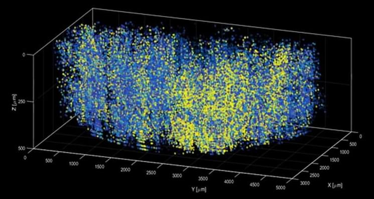
A new method – which has been dubbed “light beads microscopy” – is described in the journal Nature. This offers a creative solution that pushes the limits of imaging speed and is limited only by the physical nature of fluorescence itself. It eliminates the “deadtime” between sequential laser pulses when no neuroactivity is recorded and at the same time the need for scanning.
The technique breaks one strong pulse into 30 smaller sub-pulses, each at a different strength, which dive into 30 different depths of scattering but induce the same amount of fluorescence at each depth. This is accomplished with a cavity of mirrors that staggers the firing of each pulse in time and ensures that they can all reach their target depths via a single microscope focusing lens. Using this approach, the only limit to the rate at which samples can be recorded is the time it takes the fluorescent tags to flare. That means broad swathes of the brain can be recorded within the same time it would take a conventional two-photon microscope to capture a much smaller network of brain cells.
Scientists at Rockefeller University, New York, integrated their new system into a microscopy platform with access to a large brain volume. This enabled the recording of activity in more than a million neurons across the entire cortex of a mouse brain for the first time.
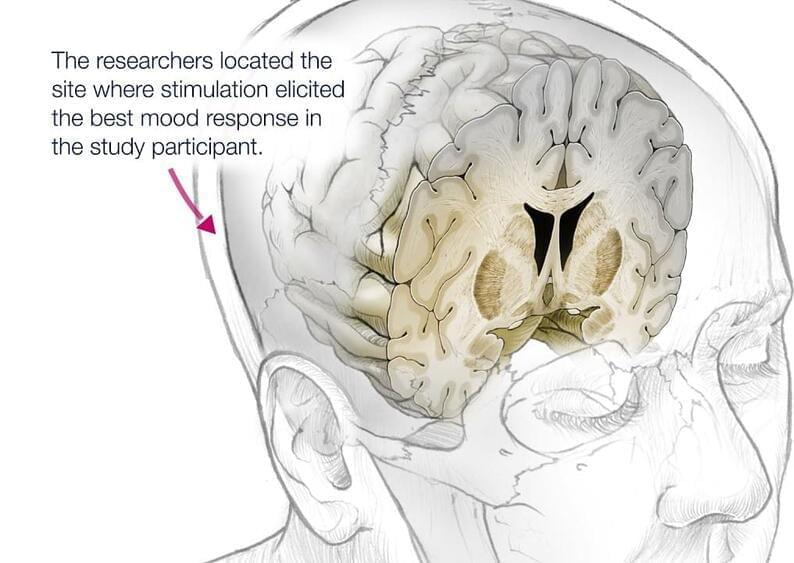
With almost instant improvement.
A team of researchers from the University of California, San Francisco Health has successfully treated a patient with severe depression by targeting the specific brain circuit involved in depressive brain patterns and resetting them thanks to a new proof-of-concept intervention.
Even though it centers around one patient, the groundbreaking study, which has now been published in Nature Medicine, is an important step toward bringing neuroscience advances and the treatment of psychiatric disorders, potentially helping millions of people who suffer from depression.
