Toggle light / dark theme

Scientists have known for decades that aerobic exercise strengthens the brain and contributes to the growth of new neurons, but few studies have examined how yoga affects the brain. A review of the science finds evidence that yoga enhances many of the same brain structures and functions that benefit from aerobic exercise.

The review, published in the journal Brain Plasticity, focused on 11 studies of the relationship between and health. Five of the studies engaged individuals with no background in yoga practice in one or more yoga sessions per week over a period of 10–24 weeks, comparing brain health at the beginning and end of the intervention. The other studies measured brain differences between individuals who regularly practice yoga and those who don’t.

Each of the studies used brain-imaging techniques such as MRI, functional MRI or single-photon emission computerized tomography. All involved Hatha yoga, which includes body movements, meditation and breathing exercises.
The idea of a cell therapy for Parkinson’s disease starts out simple: Symptoms of the progressive disease are largely driven by the deaths of dopamine-producing neurons found deep within the brain. With lower levels of the neurotransmitter come the characteristic tremors, rigidity and slow movements.

By replacing those lost nerve cells with new dopamine producers, researchers hope to renew the brain’s connection to the body’s muscles and improve a person’s overall motor function.

But in the brain, everything becomes more complicated. On top of the risk of immune system rejection that comes with any kind of living tissue transplant, it’s important to make sure the implanted cells function correctly and do not pick up any dangerous genetic mutations as they grow.

Fresh insights into damaging proteins that build up in the brains of people with Alzheimer’s disease could aid the quest for treatments.

A study in mice reveals how the two proteins work together to disrupt communication between brain cells.

Scientists observed how proteins—called amyloid beta and tau—team up to hamper key genes responsible for brain messaging. By changing how genes are expressed in the brain, the proteins can affect its normal function.

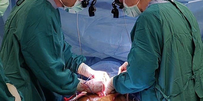
The procedure, donation after circulatory death or DCD, involves taking organs from a donor whose heart has stopped beating after being taken off of life support after a fatal injury or illness when there is no potential for recovery.

Conventional organ donations occur after brain death, which means that while all brain functions have stopped and the person is legally and clinically dead, machines can continue to keep oxygen and blood flowing throughout the body, preserving the healthy organs for donation.

After a circulatory death, however, organs are deprived of oxygen as the circulatory system shuts down, potentially damaging the donor organs and making it difficult to use them for transplant.

In mouse models of Alzheimer’s disease, the investigational drug candidates known as CMS121 and J147 improve memory and slow the degeneration of brain cells. Now, Salk researchers have shown how these compounds can also slow aging in healthy older mice, blocking the damage to brain cells that normally occurs during aging and restoring the levels of specific molecules to those seen in younger brains.

The research, published last month in the journal eLife, suggests that the drug candidates may be useful for treating a broader array of conditions and points out a new pathway that links normal aging to Alzheimer’s disease.

“This study further validated these two compounds not only as Alzheimer’s drug candidates but also as potentially more widely useful for their anti-aging effects,” says Pamela Maher, a senior staff scientist at Salk and a co-corresponding author of the new paper.

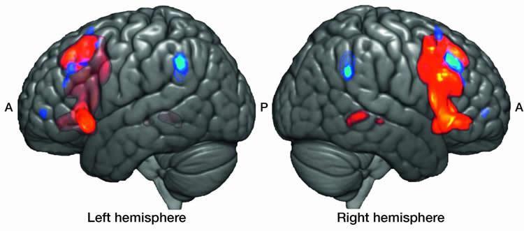
Summary: Researchers reveal the right homologue of the Broca’s area plays a major role in the processing of music.

Source: Max Planck Institute.

Vincent Cheung, along with Angela Friederici, has been investigating non-local dependencies in music and trying to determine how the human brain processes them. In language and music, dependencies are conceptual threads that bind two things together. Non-local dependencies bind non-adjacent items. For example, in pop music, the second instance of a verse, following a chorus, would have a non-local dependency with the first instance of the verse. Experientially, it is clear to us that we are hearing a sequence that we have heard before. According to Cheung, composers use such devices to build up our expectations and elicit strong emotional responses to the music. But how does the brain recognize these patterns and what does this have to do with Paul Broca?

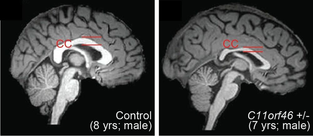
Using a targeted gene epigenome editing approach in the developing mouse brain, Johns Hopkins Medicine researchers reversed one gene mutation that leads to the genetic disorder WAGR syndrome, which causes intellectual disability and obesity in people. This specific editing was unique in that it changed the epigenome—how the genes are regulated—without changing the actual genetic code of the gene being regulated.

The researchers found that this gene, C11orf46, is an important regulator during . Specifically, it turns on and off the direction-sensing proteins that help guide the long fibers growing out of newly formed neurons responsible for sending electrical messages, helping them form into a bundle, which connects the two hemispheres of the brain. Failure to properly form this bundled structure, known as the , can lead to conditions such as , autism or other brain .

“Although this work is early, these findings suggest that we may be able to develop future epigenome editing therapies that could help reshape the neural connections in the brain, and perhaps prevent developmental disorders of the brain from occurring,” says Atsushi Kamiya, M.D., Ph.D., associate professor of psychiatry and at the Johns Hopkins University School of Medicine.

A ketone-supplemented diet may protect neurons from death during the progression of Alzheimer’s disease, according to research in mice recently published in JNeurosci.

Early in the development of Alzheimer’s disease, the brain becomes over excited, potentially through the loss of inhibitory, or GABAergic, interneurons that keep other neurons from signaling too much. Because interneurons require more energy compared to other neurons, they may be more susceptible to dying when they encounter the Alzheimer’s disease protein amyloid beta. Amyloid beta has been shown to damage mitochondria — the metabolic engine for cells — by interfering with SIRT3, a protein that preserves mitochondrial functions and protects neurons.

Cheng et al. genetically reduced levels of SIRT3 in mouse models of Alzheimer’s disease. Mice with low levels of SIRT3 experienced a much higher mortality rate, more violent seizures, and increased interneuron death compared to the mice from the standard Alzheimer’s disease model and control mice. However, the mice with reduced levels of SIRT3 experienced fewer seizures and were less likely to die when they ate a diet rich in ketones, a specific type of fatty acid. The diet also increased levels of SIRT3 in the mice.