Toggle light / dark theme

A new study by Florida State University researchers may help answer some of the most perplexing questions surrounding Alzheimer’s disease, an incurable and progressive illness affecting millions of families around the globe.

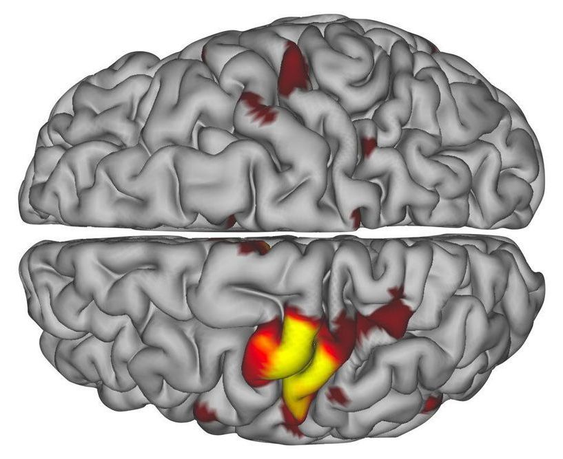
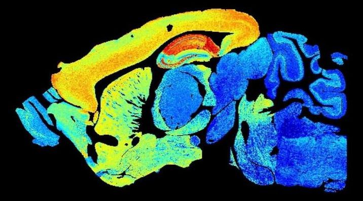
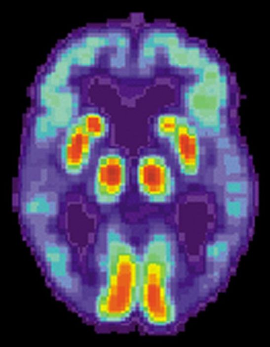
FSU Assistant Professor of Psychology Aaron Wilber and graduate student Sarah Danielle Benthem showed that the way two parts of the interact during sleep may explain symptoms experienced by Alzheimer’s patients, a finding that opens up new doors in dementia research. It is believed that these interactions during sleep allow memories to form and thus failure of this normal system in a brain of a person with Alzheimer’s disease may explain why memory is impaired.

The study, a collaboration among the FSU Program in Neuroscience, the University of California, Irvine, and the University of Lethbridge in Alberta, Canada, was published online in the journal Current Biology and will appear in the publication’s July 6 issue.
:ooooooo.


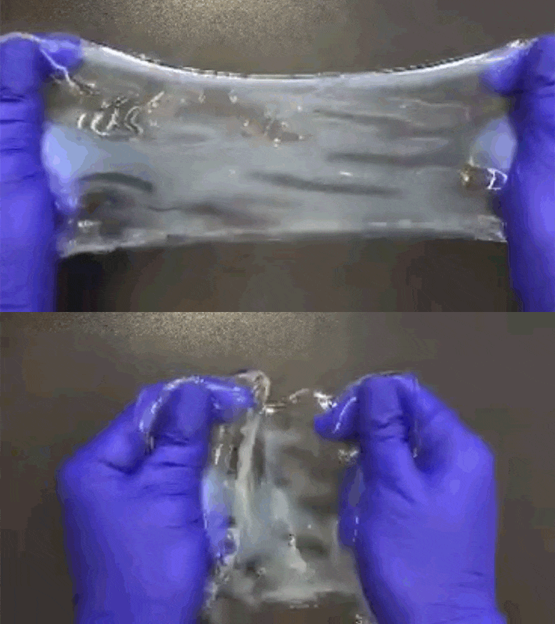
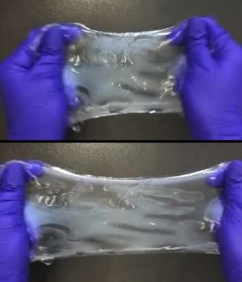
Superhero-like stretching capabilities aren’t just for Elastigirl. Researchers at the Massachusetts Institute of Technology have come up with a new technology that can make any tissue sample exceptionally flexible.

ELAST technology (Entangled Link-Augmented Stretchable Tissue-hydrogel) is a chemical process that makes tissue samples very thin, very stretchy, compressible, and borderline indestructible. With it, lab technicians can more quickly and easily conduct fluorescent labeling in cells, proteins, or other genetic materials within organs like the brain or lungs. That, in turn, could enable faster research discoveries.

The MIT researchers published their work last month in the journal Nature Methods.

How does our memory work, and how can we optimize its mechanisms on a daily basis? These questions are at the heart of many neuroscience research projects. Among the brain structures examined to better understand memory mechanisms, the reward system is now at the center of investigations. Through the examination of brain activity in healthy human subjects, scientists from the University of Geneva (UNIGE) have highlighted the lasting positive effect of a reward—monetary, in this case—on the ability of individuals to retain a variety of information. Moreover, and much more surprisingly, the research team demonstrated that the average accumulation of reward should be neither too small nor too large. By ensuring an effective neural dialog between the reward circuit and the memory circuit, this delicate balance allows the proper encoding of memories in our brain. These results can be read in Nature Communications.

Empirically, it seems quite logical that obtaining a can improve the memories associated with it. But what are the brain mechanisms at work, and how can we exploit them to optimize our memory capacity?

“The positive influence of a reward on memory is a well-known phenomenon,” says Sophie Schwartz, full professor in the Department of Basic Neurosciences at the UNIGE Faculty of Medicine, who led this work. “However, our experiment aimed to take a further step in understanding this mechanism by looking at two important aspects: does the effect last over time and what role does the accumulation of reward play?”

A neuroscientist’s neon pink arm cast led him and fellow researchers at Washington University School of Medicine in St. Louis to discover previously undetected neuronal pulses in the human brain that activate after an immobilizing illness or injury.

Just as our human relationships and connections can nudge, push, or dramatically shift societal values and consequences, the connections between neurons form intricate networks that dictate the outcome of your mind. Your thoughts, memories, behaviors; your values, world view, mental health—everything that makes you you is calculated and stored in these connections, called synapses, that dot our brains like billions of stars in the night sky.

If a connectome—a large-scale snapshot of all your neural connections—is a loose “copy” of you at one moment in time, synapses are a fluid representation of how you change and grow through time. Similar to human connections, synapses come in different varieties and evolve as we age. Yet until now, capturing how these synapses change as we move through time has been nearly impossible.

Last week, in a technological tour-de-force, a European team from the United Kingdom, France, and Sweden, led by Dr. Seth G.N. Grant at the University of Edinburgh, redefined impossibility with a paper in Science. Peering into the brains of mice at different ages—one day, one week, and all the way up to an elderly 18 months—the team constructed maps of roughly 5 billion synapses, outlining a timeline of their diversity and numbers in over 100 different brain regions with age.

Chemical process called ELAST allows labeling probes to infuse more quickly, and makes samples tough enough for repeated handling.

When there’s a vexing problem to be solved, people sometimes offer metaphorical advice such as “stretching the mind” or engaging in “flexible” thinking, but in confronting a problem facing many biomedical research labs, a team of MIT researchers has engineered a solution that is much more literal. To make imaging cells and molecules in brain and other large tissues easier while also making samples tough enough for years of handling in the lab, they have come up with a chemical process that makes tissue stretchable, compressible, and pretty much indestructible.

“ELAST” technology, described in a new paper in Nature Methods, provides scientists a very fast way to fluorescently label cells, proteins, genetic material, and other molecules within brains, kidneys, lungs, hearts, and other organs. That’s because when such tissues can be stretched out or squished down thin, labeling probes can infuse them far more rapidly. Several demonstrations in the paper show that even after repeated expansions or compressions to speed up labeling, tissues snap back to their original form unaltered except for the new labels.

A research team at Hadassah-University Medical Center in Jerusalem’s Ein Kerem has discovered what they believe causes coronavirus patients to become seriously ill and even die. They also say they have a way to treat the cause before it’s too late.

At least 30% of patients with coronavirus develop blood clots that block the flow of blood to their kidneys, heart and brain, as well as the lungs, according to international research.


Hadassah researchers discovered that the patients who form these fatal clots have an increased level of alpha defensin protein in their blood, explained Dr. Abd Alrauf Higavi, who directs a lab at Hadassah and has been studying blood clots for 30 years.

“Patients with mild symptoms have a low concentration of alpha defensin,” he said. “Patients with strong disease symptoms have high levels. The people who die have very high levels.”

The Hadassah team studied more than 700 blood samples from 80 patients who were admitted to the medical center during the first peak of the coronavirus outbreak in Israel. The results show that alpha defensin speeds up blood clot formation, which can cause pulmonary embolism, heart attacks and stroke. In addition, when blood clots form in the alveoli, whose function it is to exchange oxygen and carbon dioxide molecules to and from the bloodstream, this can lead to respiratory distress and eventually intubation.

Multiple studies have shown that around 80% of coronavirus patients who are intubated have died.

A pair of researchers at University College London has found that people with low socioeconomic status experience more declines in age-related functions as they grow older than do people who have a higher socioeconomic status. In their paper published in Proceedings of the National Academy of Sciences, Andrew Steptoe and Paola Zaninotto describe their study of data from the English Longitudinal Study of Ageing, and what they learned.

Prior research has shown that tend to suffer more adverse health effects than those who are better off. They also tend to die younger. But one area of aging that has not been well-studied is the impact of poverty on age-related functional decline, associated with such symptoms as loss of hearing or muscle strength. To learn more about the relationship between socioeconomic status and age-related functional decline, the researchers analyzed data in the English Longitudinal Study of Ageing—an ongoing long-term study of the aging process. Launched in 2002, the study involved collecting data on volunteers aged 50 and over as they grew older. The data includes both medical and physical information, along with test results designed to measure cognitive and emotional levels. The data sample for this new effort included information on 5,018 people 52 years of age or older as they aged over periods of six to eight years.

The researchers found that people living at the lower end of the economic spectrum performed worse on every measure of age-related functionality. Those less well-off lost grip strength, lung function, gait speed, processing speed and executive function. They also tended to report enjoying life less than those who were more affluent. The researchers noted their findings were independent of race, gender, education or age. They also found that those of lesser means experienced more and were more likely to be depressed.

In 2005, University of California, Berkeley, researchers made the surprising discovery that making conjoined twins out of young and old mice—such that they share blood and organs—can rejuvenate tissues and reverse the signs of aging in the old mice. The finding sparked a flurry of research into whether a youngster’s blood might contain special proteins or molecules that could serve as a “fountain of youth” for mice and humans alike.

But a new study by the same team shows that similar age-reversing effects can be achieved by simply diluting the of old mice—no needed.

In the study, the team found that replacing half of the blood plasma of old mice with a mixture of saline and albumin—where the albumin simply replaces that was lost when the original blood plasma was removed—has the same or stronger rejuvenation effects on the brain, liver and muscle than pairing with young mice or young blood exchange. Performing the same procedure on had no detrimental effects on their health.