Toggle light / dark theme

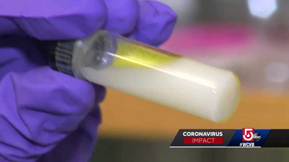
A group of scientists at Northeastern University are making progress using nanotechnology to prevent, diagnose and fight the coronavirus.

Thomas Webster, professor of chemical engineering at Northeastern University, has been working with nanotechnology for decades. Now, he and his team are finding new applications with the coronavirus.

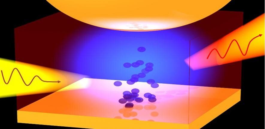
Researchers have developed a method to ‘squeeze’ visible light in order to see inside tiny memory devices. The technique will allow researchers to probe how these devices break down and how their performance can be improved for a range of applications.

The team, led by the University of Cambridge, used the technique to investigate the materials used in random access memories, while in operation. The results, reported in the journal Nature Electronics, will allow detailed study of these materials, which are used in devices.

The ability to understand how structural changes characterize the function of these materials, which are used for , ultra-responsive devices called memristors, is important to improve their performance. However, looking inside the 3D nanoscale devices is difficult using traditional techniques.

Could make awesome computers.


Materials scientists who work with nano-sized components have developed ways of working with their vanishingly small materials. But what if you could get your components to assemble themselves into different structures without actually handling them at all?

Verner Håkonsen works with cubes so tiny that nearly 5 billion of them could fit on a pinhead.

He cooks up the cubes in the NTNU NanoLab, in a weird-looking glass flask with three necks on the top using a mixture of chemicals and special soap.

Virtual reality software which allows researchers to ‘walk’ inside and analyse individual cells could be used to understand fundamental problems in biology and develop new treatments for disease.

The software, called vLUME, was created by scientists at the University of Cambridge and 3D image analysis software company Lume VR Ltd. It allows super-resolution microscopy data to be visualised and analysed in virtual reality, and can be used to study everything from individual proteins to entire cells. Details are published in the journal Nature Methods.

Super-resolution microscopy, which was awarded the Nobel Prize for Chemistry in 2014, makes it possible to obtain images at the nanoscale by using clever tricks of physics to get around the limits imposed by light diffraction. This has allowed researchers to observe molecular processes as they happen. However, a problem has been the lack of ways to visualise and analyse this data in three dimensions.

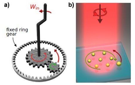
“In previous work, the researchers discovered that when optical matter is exposed to circularly polarized light, it rotates as a rigid body in the direction opposite the polarization rotation. In other words, when the incident light rotates one way the optical matter array responds by spinning the other. This is a manifestation of “negative torque”. The researchers speculated that a machine could be developed based on this new phenomenon.

In the new work, the researchers created an optical matter machine that operates much like a mechanical machine based on interlocking gears. In such machines, when one gear is turned, a smaller interlocking gear will spin in the opposite direction. The optical matter machine uses circularly polarized light from a laser to create a nanoparticle array that acts like the larger gear by spinning in the optical field. This “optical matter gear” converts the circularly polarized light into orbital, or angular, momentum that influences a nearby probe particle to orbit the nanoparticle array (the gear) in the opposite direction.”


Researchers have developed a tiny new machine that converts laser light into work. These optically powered machines self-assemble and could be used for nanoscale manipulation of tiny cargo for applications such as nanofluidics and particle sorting.

“Our work addresses a long-standing goal in the nanoscience community to create self-assembling that can perform work in conventional environments such as room temperature liquids,” said research team leader Norbert F. Scherer from the University of Chicago.

Scherer and colleagues describe the new nanomachines in Optica. The machines are based on a type of matter known as optical matter in which metal nanoparticles are held together by light rather than the that hold together the atoms that make up typical matter.
Based on optical matter, new machines could be used to move and manipulate tiny particles.

Researchers have developed a tiny new machine that converts laser light into work. These optically powered machines self-assemble and could be used for nanoscale manipulation of tiny cargo for applications such as nanofluidics and particle sorting.

“Our work addresses a long-standing goal in the nanoscience community to create self-assembling nanoscale machines that can perform work in conventional environments such as room temperature liquids,” said research team leader Norbert F. Scherer from the University of Chicago.

One of the most remarkable recent advances in biomedical research has been the development of highly targeted gene-editing methods such as CRISPR that can add, remove, or change a gene within a cell with great precision. The method is already being tested or used for the treatment of patients with sickle cell anemia and cancers such as multiple myeloma and liposarcoma, and today, its creators Emmanuelle Charpentier and Jennifer Doudna received the Nobel Prize in chemistry.

While is remarkably precise in finding and altering genes, there is still no way to target treatment to specific locations in the body. The treatments tested so far involve removing or immune system T cells from the body to modify them, and then infusing them back into a patient to repopulate the bloodstream or reconstitute an immune response—an expensive and time-consuming process.

Building on the accomplishments of Charpentier and Doudna, Tufts researchers have for the first time devised a way to directly deliver gene-editing packages efficiently across the and into specific regions of the brain, into immune system cells, or to specific tissues and organs in mouse models. These applications could open up an entirely new line of strategy in the treatment of neurological conditions, as well as cancer, infectious disease, and autoimmune diseases.