Toggle light / dark theme

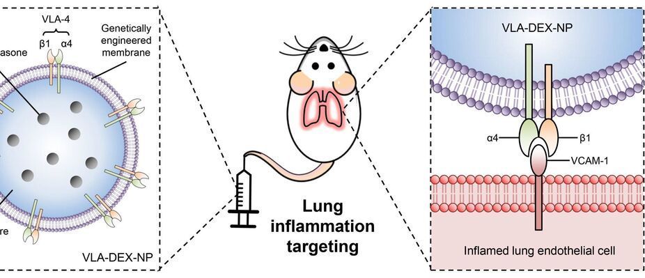
Nanoengineers at the University of California San Diego have developed immune cell-mimicking nanoparticles that target inflammation in the lungs and deliver drugs directly where they’re needed. As a proof of concept, the researchers filled the nanoparticles with the drug dexamethasone and administered them to mice with inflamed lung tissue. Inflammation was completely treated in mice given the nanoparticles, at a drug concentration where standard delivery methods did not have any efficacy.

The researchers reported their findings in Science Advances on June 16.

What’s special about these is that they are coated in a cell membrane that’s been genetically engineered to look for and bind to inflamed . They are the latest in the line of so-called cell membrane-coated nanoparticles that have been developed by the lab of UC San Diego nanoengineering professor Liangfang Zhang. His lab has previously used cell membrane-coated nanoparticles to absorb toxins produced by MRSA; treat sepsis; and train the immune system to fight cancer. But while these previous cell membranes were naturally derived from the body’s , the cell membranes used to coat this dexamethasone-filled nanoparticle were not.

Check out my short video in which I explain some super exciting research in the area of nanotechnology: de novo protein lattices! I specifically discuss a journal article by Ben-Sasson et al. titled “Design of biologically active binary protein 2D materials”.


Here, I explain an exciting nanotechnology paper “Design of biologically active binary protein 2D materials” (https://doi.org/10.1038/s41586-020-03120-8).

Though I am not involved in this particular research myself, I have worked in adjacent areas such as de novo engineering of aggregating antimicrobial peptides, synthetic biology, nanotechnology-based tools for neuroscience, and gene therapy. I am endlessly fascinated by this kind of computationally driven de novo protein design and would love to incorporate it in my own research at some point in the future.

I am a PhD candidate at Washington University in St. Louis and the CTO of the startup company Conduit Computing. I am also a published science fiction writer and a futurist. To learn more about me, check out my website: https://logancollinsblog.com/.

Circa 2015


Coatings that attract water (hydrophilic) are useful for anti-fogging applications6; any liquid water spreads out into a thin film thereby maintaining transparency. This is more favorable than using hydrophobic surfaces for anti-fogging as this requires a surface to be tilted for the droplets to roll off and transparency be maintained. Hydrophilic surfaces can also be used for self-cleaning7. Previous examples of superhydrophilic surfaces include the use of polymer–nanoparticle coatings8,9,10,11 however mechanical durability was not investigated.

Coatings with surface tensions lower than that of water (72 mN m–1) but higher than that of oils12 (20–30 mN m–1) will attract oils (oleophilic) but repel water and can be used to create oil–water separators13,14,15. When applied to a porous substrate, the coating will allow the passage of oil but block the passage of water, resulting in their separation. In addition, their water repellency also makes them ideal for self-cleaning4,16 and anti-icing17,18,19 applications. Anti-icing surfaces are typically superhydrophobic as supercooled droplets of water are able to roll off the cold surface before freezing and any ice formed is weakly adhered compared to hydrophilic surfaces due to an air cushion18,20.

Coatings with lower surface tensions (∼ 20 mN m–1 or less) will repel both oil (oleophobic) and water and are useful for anti-fouling such as in medical and transport applications, where both the oil-repellency and nanostructuring are of importance21,22,23,24,25,26,27. Previous work was not suitable for such applications as either the durability28 or oil-repellency29 was not optimal. The oil repellency also makes these surfaces ideal for anti-smudge applications30,31 where the oils from fingers are not deposited onto the surface and the surface remains clear. The water repellency means these coatings can also be used in self-cleaning and anti-icing applications.

Circa 2019


The evolution of micro and nanofabrication approaches significantly spurred the advancements of cardiac tissue engineering over the last decades. Engineering in the micro and nanoscale allows for the rebuilding of heart tissues using cardiomyocytes. The breakthrough of human induced pluripotent stem cells expanded this field rendering the development of human tissues from adult cells possible, thus avoiding the ethical issues of the usage of embryonic stem cells but also creating patient-specific human engineered tissues. In the case of the heart, the combination of cardiomyocytes derived from human induced pluripotent stem cells and micro/nano engineering devices gave rise to new therapeutic approaches of cardiac diseases. In this review, we survey the micro and nanofabrication methods used for cardiac tissue engineering, ranging from clean room-based patterning (such as photolithography and plasma etching) to electrospinning and additive manufacturing. Subsequently, we report on the main approaches of microfluidics for cardiac culture systems, the so-called “Heart on a Chip”, and we assess their efficacy for future development of cardiac disease modeling and drug screening platforms.

German nanotechnology specialist attocube says its attoDRY800 cryostat enables quantum scientists to “reclaim the optical table” and focus on their research not the experimental set-up.

Twin-track innovations in cryogenic cooling and optical table design are “creating the space” for fundamental scientific breakthroughs in quantum communications, allowing researchers to optimize the performance of secure, long-distance quantum key distribution (QKD) using engineered single-photon-emitting light sources.

In a proof-of-concept study last year, Tobias Heindel and colleagues in the Institute of Solid State Physics at the Technische Universität (TU) Berlin, Germany, implemented a basic QKD testbed in their laboratory. The experimental set-up uses a semiconductor quantum-dot emitter to send single-photon pulses along an optical fibre to a four-port receiver that analyses the polarization state of the transmitted qubits.

Circa 2020 o,.o!


Every robot is, at its heart, a computer that can move. That is true from the largest plane-sized flying machines down to the smallest of controllable nanomachines, small enough to someday even navigate through blood vessels.

New research, published August 26 in Nature, shows that it is possible to build legs into robots mere microns in length. When powered by lasers, these tiny machines can move, and some day, they may save lives in operating rooms or even, possibly, on the battlefield.

This project, funded in part by the Army Research Office and the Air Force Office of Scientific Research, demonstrated that, adapting principles from origami, nano-scale legged robots could be printed and then directed.

Researchers from Cornell University’s School of Applied and Engineering Physics and Samsung’s Advanced Institute of Technology have created a first-of-its-kind metalens—a metamaterial lens—that can be focused using voltage instead of mechanically moving its components.

The proof of concept opens the door to a range of compact varifocal lenses for possible use in many imaging applications such as satellites, telescopes and microscopes, which traditionally focus light using curved lenses that adjust using mechanical parts. In some applications, moving traditional glass or plastic lenses to vary the focal distance is simply not practical due to space, weight or size considerations.

Metalenses are flat arrays of nano-antennas or resonators, less than a micron thick, that act as focusing devices. But until now, once a metalens was fabricated, its was hard to change, according to Melissa Bosch, doctoral student and first author of a paper detailing the research in the American Chemical Society’s journal Nano Letters.

MIT engineers have discovered a way to generate electricity using tiny carbon particles that can create an electric current simply by interacting with an organic solvent in which they’re floating. The particles are made from crushed carbon nanotubes (blue) coated with a Teflon-like polymer (green). Credit: Jose-Luis Olivares, MIT. Based on a figure courtesy of the researchers.

A new material made from carbon nanotubes can generate electricity by scavenging energy from its environment.

MIT engineers have discovered a new way of generating electricity using tiny carbon particles that can create a current simply by interacting with liquid surrounding them.

MIT engineers have discovered a new way of generating electricity using tiny carbon particles that can create a current simply by interacting with liquid surrounding them.

The liquid, an , draws electrons out of the particles, generating a current that could be used to drive or to power micro-or nanoscale robots, the researchers say.

“This mechanism is new, and this way of generating is completely new,” says Michael Strano, the Carbon P. Dubbs Professor of Chemical Engineering at MIT. “This technology is intriguing because all you have to do is flow a solvent through a bed of these particles. This allows you to do electrochemistry, but with no wires.”