Toggle light / dark theme

Luv this.


MIT biological engineers have devised a way to record complex histories in the DNA of human cells, allowing them to retrieve “memories” of past events, such as inflammation, by sequencing the DNA.

This analog memory storage system—the first that can record the duration and/or intensity of events in human cells—could also help scientists study how cells differentiate into various tissues during embryonic development, how cells experience environmental conditions, and how they undergo genetic changes that lead to disease.

“To enable a deeper understanding of biology, we engineered human cells that are able to report on their own history based on genetically encoded recorders,” says Timothy Lu, an associate professor of electrical engineering and computer science, and of biological engineering. This technology should offer insights into how gene regulation and other events within cells contribute to disease and development, he adds.

Read more

Many lower organisms retain the miraculous ability to regenerate form and function of almost any tissue after injury. Humans share many of our genes with these organisms, but our capacity for regeneration is limited. Scientists at the MDI Biological Laboratory in Bar Harbor, Maine, are studying the genetics of these organisms to find out how regenerative mechanisms might be activated in humans.

The ability of animals to regenerate body parts has fascinated scientists since the time of Aristotle. But until the advent of sophisticated tools for genetic and computational analysis, scientists had no way of studying the genetic machinery that enables regeneration. Using such tools, scientists at the MDI Biological Laboratory have identified genetic regulators governing regeneration that are common across species.

In a paper published in the journal PLOS ONE, MDI Biological Laboratory scientists Benjamin L. King, Ph.D., and Voot P. Yin, Ph.D., identified these common genetic regulators in three regenerative species: the zebrafish, a common aquarium fish originally from India; the axolotl, a salamander native to the lakes of Mexico; and the bichir, a ray-finned fish from Africa.

Read more

Abstract.

Developmental dyslexia is a common disorder with a strong genetic component, but the underlying molecular mechanisms are still unknown. Several candidate dyslexia-susceptibility genes, including KIAA0319, DYX1C1, and DCDC2, have been identified in humans. RNA interference experiments targeting these genes in rat embryos have shown impairments in neuronal migration, suggesting that defects in radial cortical migration could be involved in the disease mechanism of dyslexia. Here we present the first characterisation of a Kiaa0319 knockout mouse line. Animals lacking KIAA0319 protein do not show anatomical abnormalities in any of the layered structures of the brain. Neurogenesis and radial migration of cortical projection neurons are not altered, and the intrinsic electrophysiological properties of Kiaa0319 -deficient neurons do not differ from those of wild-type neurons. Kiaa0319 overexpression in cortex delays radial migration, but does not affect final neuronal position. However, knockout animals show subtle differences suggesting possible alterations in anxiety-related behaviour and in sensorimotor gating. Our results do not reveal a migration disorder in the mouse model, adding to the body of evidence available for Dcdc2 and Dyx1c1 that, unlike in the rat in utero knockdown models, the dyslexia-susceptibility candidate mouse homolog genes do not play an evident role in neuronal migration. However, KIAA0319 protein expression seems to be restricted to the brain, not only in early developmental stages but also in adult mice, indicative of a role of this protein in brain function. The constitutive and conditional knockout lines reported here will be useful tools for further functional analyses of Kiaa0319.

Read more

(credit: NIH National Institute on Aging)

Anti-inflammatory drug mefenamic acid completely reversed memory loss and brain inflammation in mice genetically engineered to develop symptoms of Alzheimer’s disease and amyloid beta-induced memory loss, a team led by David Brough, PhD, from the University of Manchester has discovered.

The non-steroidal anti-inflammatory drug (NSAID) drug targets an important inflammatory pathway called the NLRP3 inflammasome, which damages brain cells, according to Brough. This is the first time a drug has been shown to target this inflammatory pathway, highlighting its importance in the disease model, Brough said.

Read more

By Kevin Kang

A recent article in ScienceDaily reviews a new approach in Synthetic Biology that allows cells to respond to a series of input stimuli and simultaneously remember the order of these stimuli over many generations. As noted by the senior investigator, Timothy Lu from MIT, combining computation with memory creates complex cellular circuits that can perform logic functions and store memories of events by encoding them in their DNA (1,2). In their current work, Dr. Lu and his colleagues created cells that can remember and respond to three different inputs, including chemical signals in a particular order, and in the future may be able to incorporate even more inputs (1,2,3). The cellular machines thus created are referred to as biological “state machines” because they exist in different states depending on the identity and order of inputs that they receive. The state machines rely on enzymes called recombinases. When activated by a specific input, recombinases either delete or invert a particular segment of DNA depending on the orientation of two DNA target sequences known as recognition sites. The segment of DNA between these sites may have recognition sites for other recombinases that respond to different inputs. Flipping or deleting these sites permanently changes what will happen if a second or third recombinase is later activated. Therefore, a cell’s history is determined by sequencing its DNA. In a version of this system with just two inputs, there are five possible states for this circuit: states corresponding to no input, input A alone, input B alone, A followed by B, and B followed by A. Dr. Lu’s team in MIT has designed and built circuits that record up to three inputs, in which sixteen states are possible (1,2).

Besides creating circuits that record events in a cell’s life and then transmit these memories to future generations, the researchers from MIT also placed genes into the array of recombinase binding sites along with genetic regulatory elements. In these circuits, when recombinases rearrange the DNA, the circuits record the information as well as control which genes get turned on and off. Lu’s lab tested this work in bacteria by color coding the identity and order of input stimuli, so input A followed by B would would lead to bacteria fluorescing red and green, but input B followed by A would lead to red and blue fluorescence. Hence, these techniques can be used not only to record the states that the cells experience over time, but also to deploy in state-dependent gene expression programs (1,2).

Read more
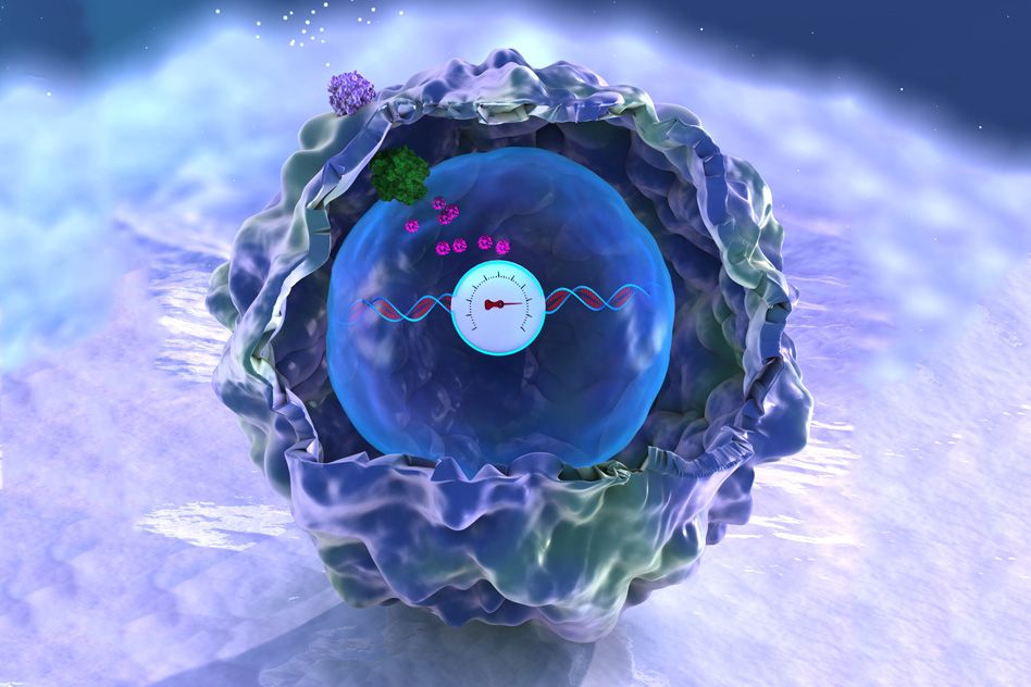
They discovered genetically engineered bacteria’s response to shocks.

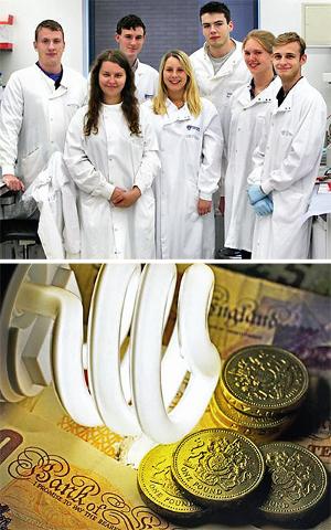
Genetically engineered E coli bacteria responds to electric shocks by producing a fluorescent protein that can be used as a light source. A team of undergraduate scientists at Newcastle University have created a lightbulb made up of living matter.

Like any other electric bulb, the living light glows to illuminate a room, but is made by replacing some of the traditional electric components in a lightbulb circuit with biological parts.

Read more

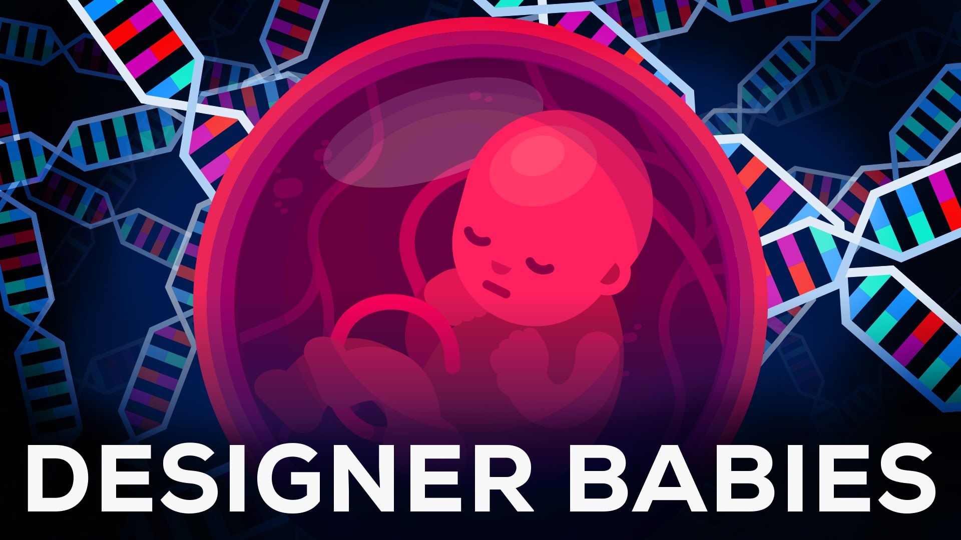
Designer babies, the end of diseases, genetically modified humans that never age. Outrageous things that used to be science fiction are suddenly becoming reality. The only thing we know for sure is that things will change irreversibly.

Support us on Patreon so we can make more videos (and get cool stuff in return): https://www.patreon.com/Kurzgesagt?ty=h

Kurzgesagt merch here: http://bit.ly/1P1hQIH

Get the music of the video here:

Soundcloud: http://bit.ly/2aRxNZd

Bandcamp: http://bit.ly/2berrSW

Read more

Leiden theoretical physicists have proven that DNA mechanics, in addition to genetic information in DNA, determines who we are. Helmut Schiessel and his group simulated many DNA sequences and found a correlation between mechanical cues and the way DNA is folded. They have published their results in PLoS One.

When James Watson and Francis Crick identified the structure of DNA molecules in 1953, they revealed that DNA information determines who we are. The sequence of the letters G, A, T and C in the famous double helix determines what proteins are made ny our cells. If you have brown eyes, for example, this is because a series of letters in your DNA encodes for proteins that build brown eyes. Each cell contains the exact same letter sequence, and yet every organ behaves differently. How is this possible?

Read more

Researchers have used CRISPR—a revolutionary new genetic engineering technique—to convert cells isolated from mouse connective tissue directly into neuronal cells.

In 2006, Shinya Yamanaka, a professor at the Institute for Frontier Medical Sciences at Kyoto University at the time, discovered how to revert adult , called fibroblasts, back into immature stem cells that could differentiate into any cell type. These so-called induced won Yamanaka the Nobel Prize in medicine just six years later for their promise in research and medicine.

Since then, researchers have discovered other ways to convert cells between different types. This is mostly done by introducing many of “master switch” genes that produce proteins that turn on entire genetic networks responsible for producing a particular cell type.

Read more