Category: genetics
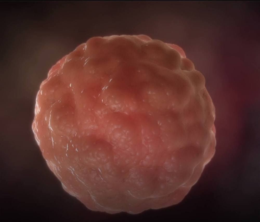
Researchers Engineer Cancer Cells To Fight Their Own Kind
Cancer cells genetically engineered to fight their own kind.

Magnetic Fields Encourage Cellular Reprogramming
Could be used in a portable device to genetically reprogram ones body.
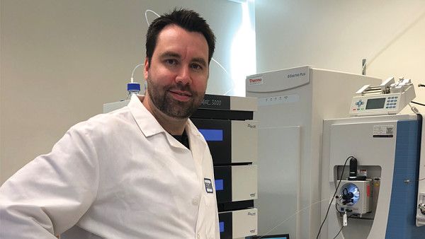
Environmental conditions, such as heat, acidity, and mechanical forces, can affect the behavior of cells. Some biologists have even shown that magnetic fields can influence them. Now, for the first time, an international team reports that low-strength magnetic fields may foster the reprogramming of cellular development, aiding in the transformation of adult cells into pluripotent stem cells (ACS Nano 2014, DOI: 10.1021/nn502923s). If confirmed, the phenomenon could lead to new tools for bioengineers to control cell fates and help researchers understand the potential health effects of changing magnetic fields on astronauts.
Biologists have been building up evidence that magnetic fields affect living things, says Michael Levin, director of Tufts University’s Center for Regenerative & Developmental Biology, who was not involved in the new study. For example, plants and amphibian embryos develop abnormally when shielded from Earth’s geomagnetic field. And there’s some clinical evidence that particular electromagnetic frequencies promote bone fracture healing and wound repair (Eur. Cytokine Network 2013, DOI: 10.1684/ecn.2013.0332).
“It’s been a huge unknown how a cell senses electromagnetic fields and then translates that into a change in identity or a change in gene expression,” says Christopher J. Lengner, a cell biologist at the University of Pennsylvania. He worked with a group of bioengineers led by Jongpil Kim of Dongguk University, in Seoul, South Korea, to see if these fields could influence a process they were all interested in: reprogramming a cell’s developmental state.
Is multiple sclerosis linked to childhood viral infections?
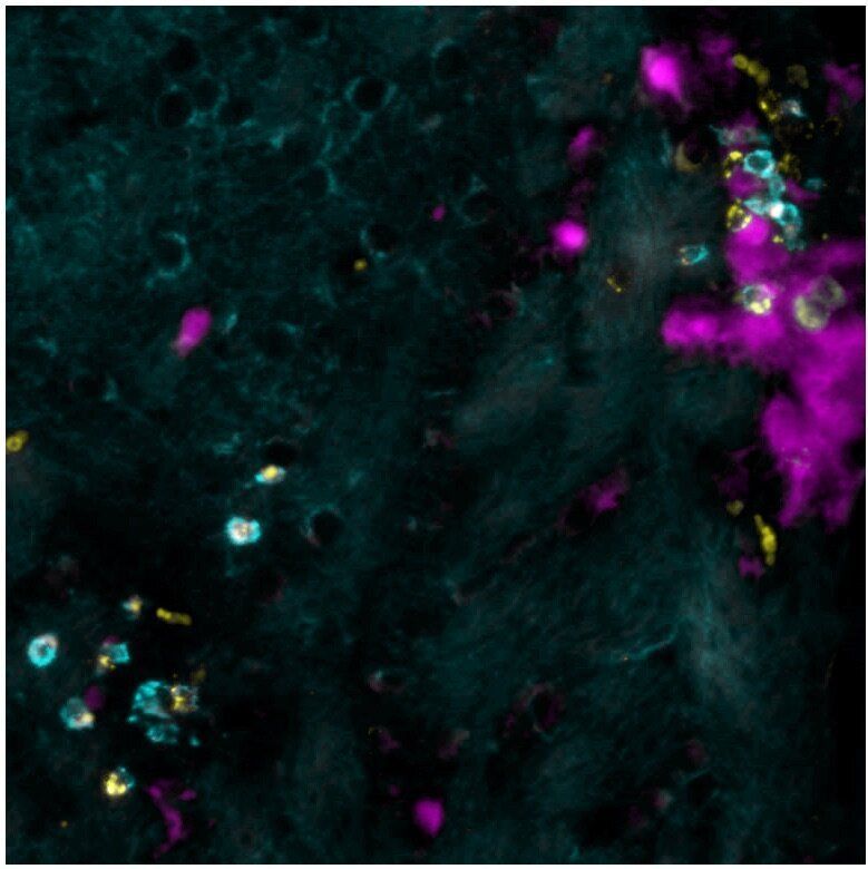
Although the exact causes of multiple sclerosis still remain unknown, it is assumed that the disease is triggered by a combination of genetic and environmental risk factors. But which? In a mouse model of the disease, researchers at the University of Geneva (UNIGE) and the Geneva University Hospitals (HUG), Switzerland, studied the potential link between transient cerebral viral infections in early childhood and the development of this cerebral autoimmune disease later in life. Indeed, the brain area affected by viral infection during childhood undergoes a change that can call, a long time later, on the immune system to turn against itself at this precise location, triggering autoimmune lesions. These results, which are published in the journal Science Translational Medicine, provide a first step in answering one of the possible environmental causes of this serious disease.
Multiple sclerosis affects one in 1,000 people in Switzerland, two-thirds of whom are women. It is the most common auto-immune disease affecting the brain. Up to date, there is still neither a cure available, nor a clear understanding of the factors that trigger this disease at around 30 years of age. “We asked ourselves whether brain viral infections that could be contracted in early childhood were among the possible causes,” says Doron Merkler, a professor in the Department of Pathology and Immunology in UNIGE’s Faculty of Medicine and senior consultant in the Clinical Pathology Service of the HUG. Such transient brain infections can be controlled quickly by the immune system, without the affected individual even noticing any symptoms. “But these transient infections may, under certain circumstances, leave a local footprint, an inflammatory signature, in the brain,” continues the researcher.

Functional hair follicles grown from stem cells
Scientists from Sanford Burnham Prebys have created natural-looking hair that grows through the skin using human induced pluripotent stem cells (iPSCs), a major scientific achievement that could revolutionize the hair growth industry. The findings were presented today at the annual meeting of the International Society for Stem Cell Research (ISSCR) and received a Merit Award. A newly formed company, Stemson Therapeutics, has licensed the technology.
More than 80 million men, women and children in the United States experience hair loss. Genetics, aging, childbirth, cancer treatment, burn injuries and medical disorders such as alopecia can cause the condition. Hair loss is often associated with emotional distress that can reduce quality of life and lead to anxiety and depression.
“Our new protocol described today overcomes key technological challenges that kept our discovery from real-world use,” says Alexey Terskikh, Ph.D., an associate professor in Sanford Burnham Prebys’ Development, Aging and Regeneration Program and the co-founder and chief scientific officer of Stemson Therapeutics. “Now we have a robust, highly controlled method for generating natural-looking hair that grows through the skin using an unlimited source of human iPSC-derived dermal papilla cells. This is a critical breakthrough in the development of cell-based hair-loss therapies and the regenerative medicine field.”

Epigenetic Reprogramming in Mammalian Development
Circa 2001
DNA methylation is a major epigenetic modification of the genome that regulates crucial aspects of its function. Genomic methylation patterns in somatic differentiated cells are generally stable and heritable. However, in mammals there are at least two developmental periods—in germ cells and in preimplantation embryos—in which methylation patterns are reprogrammed genome wide, generating cells with a broad developmental potential. Epigenetic reprogramming in germ cells is critical for imprinting; reprogramming in early embryos also affects imprinting. Reprogramming is likely to have a crucial role in establishing nuclear totipotency in normal development and in cloned animals, and in the erasure of acquired epigenetic information. A role of reprogramming in stem cell differentiation is also envisaged.
DNA methylation is one of the best-studied epigenetic modifications of DNA in all unicellular and multicellular organisms. In mammals and other vertebrates, methylation occurs predominantly at the symmetrical dinucleotide CpG (1–4). Symmetrical methylation and the discovery of a DNA methyltransferase that prefers a hemimethylated substrate, Dnmt1 , suggested a mechanism by which specific patterns of methylation in the genome could be maintained. Patterns imposed on the genome at defined developmental time points in precursor cells could be maintained by Dnmt1, and would lead to predetermined programs of gene expression during development in descendants of the precursor cells (5, 6). This provided a means to explain how patterns of differentiation could be maintained by populations of cells. In addition, specific demethylation events in differentiated tissues could then lead to further changes in gene expression as needed.
Neat and convincing as this model is, it is still largely unsubstantiated. While effects of methylation on expression of specific genes, particularly imprinted ones and some retrotransposons , have been demonstrated in vivo, it is still unclear whether or not methylation is involved in the control of gene expression during normal development (9–13). Although enzymes have been identified that can methylate DNA de novo (Dnmt3a and Dnmt3b) (14), it is unknown how specific patterns of methylation are established in the genome. Mechanisms for active demethylation have been suggested, but no enzymes have been identified that carry out this function in vivo (15–17). Genomewide alterations in methylation—brought about, for example, by knockouts of the methylase genes—result in embryo lethality or developmental defects, but the basis for abnormal development still remains to be discovered (7, 14).
Scientists discover molecular key to how cancer spreads
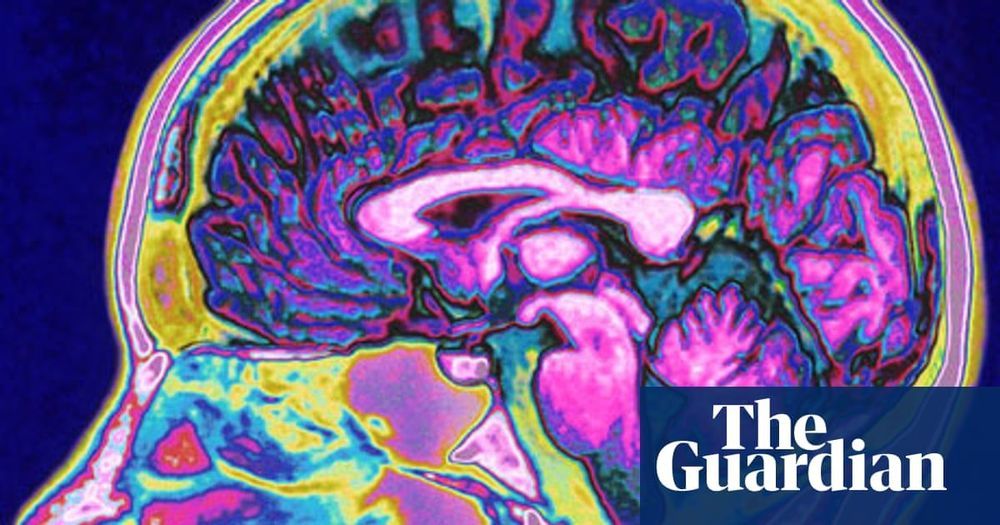
Yale researchers have discovered how metastasis, the spread of cancer cells throughout the body, is triggered on the molecular level, and have developed a tool with the potential to detect those triggers in patients with certain cancers. The discovery could lead to new ways for treating cancer.
The study was led by Andre Levchenko, the John C. Malone Professor of Biomedical Engineering and director of the Yale Systems Biology Institute at Yale’s West Campus. It was published June 26 in the journal Nature Communications. Levchenko is a member of the Yale Cancer Center.
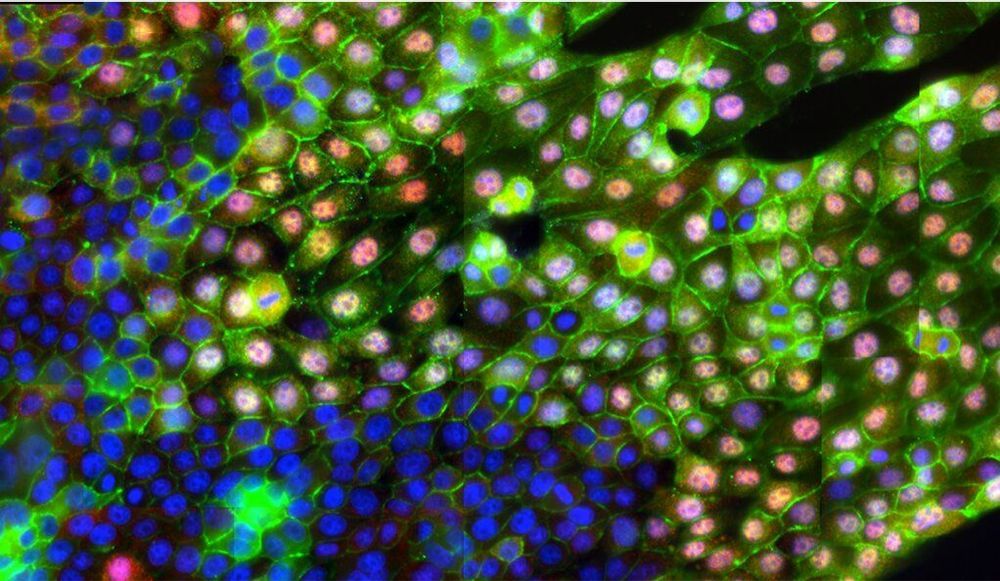
One way metastasis occurs is through epithelial-mesenchymal transition (EMT), a process that breaks neighboring cells apart from each other and sets them in motion. It’s been long assumed that chemical signals or genetic changes in the cells trigger EMT. But Levchenko’s research team found that it could be caused by a simple change in the texture of the extracellular matrix (ECM), which acts as a scaffold for cells. They discovered that an alignment of the matrix’s fibers (a common biological occurrence) can trigger the EMT process without or other stimuli.