Toggle light / dark theme

The Nobel Prize in Chemistry has been awarded to Emmanuelle Charpentier and Jennifer A. Doudna for the development of a method for genome editing.


Emmanuelle Charpentier (left), and Jennifer Doudna, are the sixth and seventh women to win the prestigious award.

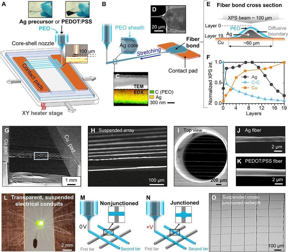
Scalability and device integration have been prevailing issues limiting our ability in harnessing the potential of small-diameter conducting fibers. We report inflight fiber printing (iFP), a one-step process that integrates conducting fiber production and fiber-to-circuit connection. Inorganic (silver) or organic {PEDOT: PSS [poly(3,4-ethylenedioxythiophene) polystyrene sulfonate]} fibers with 1- to 3-μm diameters are fabricated, with the fiber arrays exhibiting more than 95% transmittance (350 to 750 nm). The high surface area–to–volume ratio, permissiveness, and transparency of the fiber arrays were exploited to construct sensing and optoelectronic architectures. We show the PEDOT: PSS fibers as a cell-interfaced impedimetric sensor, a three-dimensional (3D) moisture flow sensor, and noncontact, wearable/portable respiratory sensors. The capability to design suspended fibers, networks of homo cross-junctions and hetero cross-junctions, and coupling iFP fibers with 3D-printed parts paves the way to additive manufacturing of fiber-based 3D devices with multilatitude functions and superior spatiotemporal resolution, beyond conventional film-based device architectures.

Small-diameter conducting fibers have unique morphological, mechanical, and optical properties such as high aspect ratio, low bending stiffness, directionality, and transparency that set them apart from other classes of conducting, film-based micro/nano structures (1–3). Orderly assembling of thin conducting fibers into an array or three-dimensional (3D) structures upscales their functional performance for device coupling. Developing new strategies to control rapid synthesis, patterning, and integration of these conducting elements into a device architecture could mark an important step in enabling new device functions and electronic designs (4, 5). To date, conducting micro/nanoscaled fibers have been produced and assembled in a number of ways, from transferring of chemically grown nanofibers/wires (6, 7), writing electrohydrodynamically deposited lines (8, 9), to drawing ultralong fibers (10, 11), wet spinning of fibers (12–14), and 2D/3D direct printing (15–18).

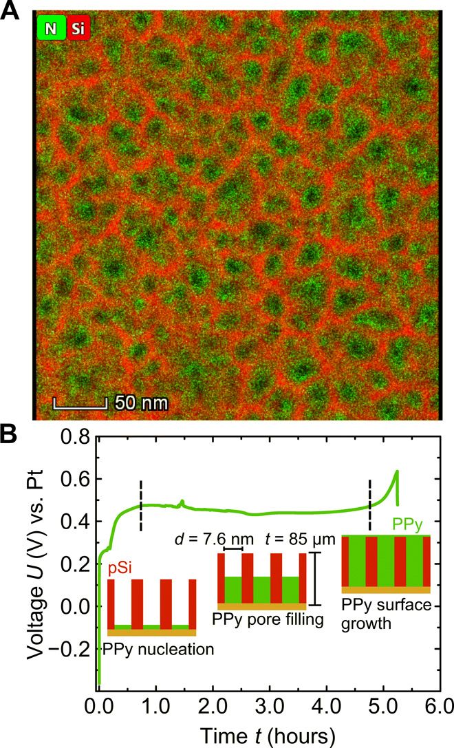
The absence of piezoelectricity in silicon makes direct electromechanical applications of this mainstream semiconductor impossible. Integrated electrical control of the silicon mechanics, however, would open up new perspectives for on-chip actuorics. Here, we combine wafer-scale nanoporosity in single-crystalline silicon with polymerization of an artificial muscle material inside pore space to synthesize a composite that shows macroscopic electrostrain in aqueous electrolyte. The voltage-strain coupling is three orders of magnitude larger than the best-performing ceramics in terms of piezoelectric actuation. We trace this huge electroactuation to the concerted action of 100 billions of nanopores per square centimeter cross section and to potential-dependent pressures of up to 150 atmospheres at the single-pore scale. The exceptionally small operation voltages (0.4 to 0.9 volts), along with the sustainable and biocompatible base materials, make this hybrid promising for bioactuator applications.

An electrochemical change in the oxidation state of polypyrrole (PPy) can increase or decrease the number of delocalized charges in its polymer backbone (1). Immersed in an electrolyte, this is also accompanied by a reversible counter-ion uptake or expulsion and thus with a marcroscopic contraction or swelling under electrical potential control, making PPy one of the most used artificial muscle materials (15).

Here, we combine this actuator polymer with the three-dimensional (3D) scaffold structure of nanoporous silicon (68) to design, similarly as found in many multiscale biological composites in nature (9), a material with embedded electrochemical actuation that consists of a few light and abundant elemental constituents (i.e., H, C, N, O, Si, and Cl).

One of the greatest mysteries in our Universe is right here on our own doorstep. No, closer — it’s in every fibre of our being.

At least 3.7 billion years ago, a few simple molecules worked together to create something new. Then a few more. And, somehow, these snowballing combinations eventually produced the first very basic living organisms that would evolve and branch out to become all life on Earth.

We don’t know the order it happened in; heck, we don’t even know when or where it happened. But new research is showing us the possibilities.

On a blustery winter day last December, a car carrying radioactive material approached one of the Port Authority of New York and New Jersey’s major transportation hubs. As the car got closer, an alarm flashed and sounded on a large monitor in the police operations center, identifying on a digital map the exact location of the vehicle and the specific radioactive isotope radiating from the car – Cesium-137. Within minutes, officers in the Port Authority Police Department – equipped with vehicle-mounted and pocket-sized radiation sensors displaying the same real-time digital map – tracked the vehicle and apprehended the suspects in a parking lot. Thankfully, the potential terrorists and radiation-emitting isotope were not a threat, as the scenario was only a drill.

The December exercise marked the capstone for DARPA’s SIGMA program, culminating a five-year effort to develop and deploy an automated, high-performance, networked radiation detection capability for counterterrorism and continuous city-to-region scale radiological and nuclear threat monitoring. The transition of the radiation-detection system took place prior to the coronavirus disease (COVID-19) pandemic. In the eight months since the SIGMA transition, DARPA has been developing and testing additional sensors under its SIGMA+ effort to detect chemical, biological and explosive threats as well.

“We want to thank the Port Authority for their outstanding support throughout the SIGMA program and their continued support as we test SIGMA+ sensors,” said Mark Wrobel, DARPA program manager in the Defense Sciences Office. “Being able to test and refine the system in the country’s largest metropolitan region was invaluable in taking SIGMA from a research project to an operationally deployed system in just five years.”

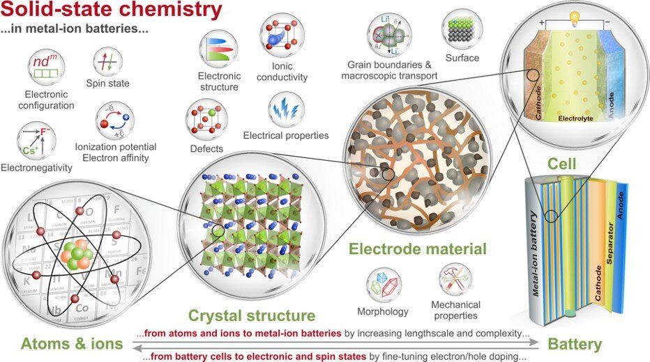
Professors from the Skoltech Center for Energy Science and Technology (CEST), Lomonosov Moscow State University and College de France shared their vision on the importance of solid state chemistry in advancements currently awaited from contemporary and prospective metal-ion batteries. The opinion was contributed as an invited review to Nature Communications.

Metal-ion batteries are the main drivers enabling a smooth transfer to renewables and green energy for a sustainable planet. The artfully designed electrode materials have greatly contributed to the development of high-performance Li-ion batteries that was eventually hallmarked by the 2019 Nobel Prize, which had signified the role solid state chemistry. Targeted design of novel metal-ion battery materials to bring the technology to the next level clearly stands as a great challenge for today’s chemistry community.

The individual properties of atoms and ions encoded in the Periodic Table along with the fundamental trends and principles multiplied by further levels of complexity constitute multitude of possible combinations for scientists to find new battery electrodes. Obviously, the researchers need solid guidelines while searching through this huge parameter space for the best chemical combinations and structures.

The bacteria cocktail eats plastic six times faster than any other bug.


A newly discovered “super-enzyme” could finally mean effective recycling of plastic bottles and other materials, scientists say. The plastic-eating bacteria can digest plastic six times faster than current methods of chemically breaking it down.

DIVE DEEPER. Read best-in-class military features and get unlimited access to Pop Mech, starting now.

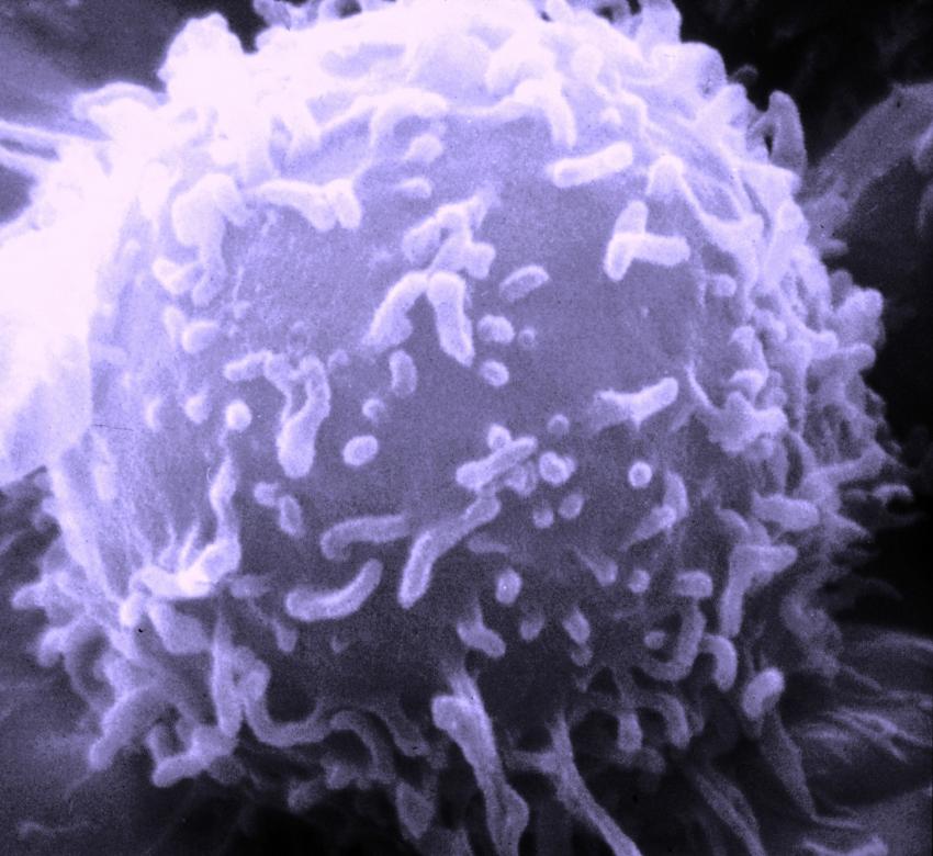
One of the key challenges in developing effective, targeted cancer treatments is the heterogeneity of the cancer cells themselves. This variation makes it difficult for the immune system to recognize, respond to and actively fight against tumors. Now, however, new advances in nanotechnology are making it possible to deliver targeted, personalized “vaccines” to treat cancer.

A new study, published on October 2, 2020 in Science Advances, demonstrates the use of charged nanoscale metal-organic frameworks for generating free radicals using X-rays within tumor tissue to kill directly. Furthermore, the same frameworks can be used for delivering immune signaling molecules known as PAMPs to activate the immune response against . By combining these two approaches into one easily administered “vaccine,” this new technology may provide the key to better local and systemic treatment of difficult-to-treat cancers.

In a collaboration between the Lin Group in the University of Chicago Department of Chemistry and the Weichselbaum Lab at University of Chicago Medicine, the research team combined expertise from inorganic chemistry and to tackle the challenging problem of properly targeting and activating an innate immune response against . This work leveraged the unique properties of nanoscale metal-organic frameworks, or nMOFs —nanoscale structures built of repeating units in a lattice formation that are capable of infiltrating tumors.

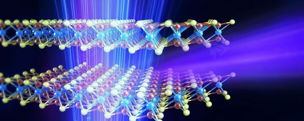
Technion researchers have developed accurate radiation sources that are expected to lead to breakthroughs in medical imaging and other areas. They have developed precise radiation sources that may replace the expensive and cumbersome facilities currently used for such tasks. The suggested apparatus produces controlled radiation with a narrow spectrum that can be tuned with high resolution, at a relatively low energy investment. The findings are likely to lead to breakthroughs in a variety of fields, including the analysis of chemicals and biological materials, medical imaging, X-ray equipment for security screening, and other uses of accurate X-ray sources.

Published in the journal Nature Photonics, the study was led by Professor Ido Kaminer and his master’s student Michael Shentcis as part of a collaboration with several research institutes at the Technion: the Andrew and Erna Viterbi Faculty of Electrical Engineering, the Solid State Institute, the Russell Berrie Nanotechnology Institute (RBNI), and the Helen Diller Center for Quantum Science, Matter and Engineering.

The researchers’ paper shows an experimental observation that provides the first proof-of-concept for theoretical models developed over the last decade in a series of constitutive articles. The first article on the subject also appeared in Nature Photonics. Written by Prof. Kaminer during his postdoc at MIT, under the supervision of Prof. Marin Soljacic and Prof. John Joannopoulos, that paper presented theoretically how two-dimensional materials can create X-rays. According to Prof. Kaminer, “that article marked the beginning of a journey towards sources based on the unique physics of two-dimensional materials and their various combinations—heterostructures. We have built on the theoretical breakthrough from that article to develop a series of follow-up articles, and now, we are excited to announce the first experimental observation on the creation of X-ray radiation from such materials, while precisely controlling the radiation parameters.”

Researchers have identified a bio-chemical circuit that supports neuron-microglia communication. When neurons are active, they release ATP. Microglia sense extracellular ATP and the compound draws the immune cell toward the neuron.circuit that supports neuron-microglia communication. When neurons are active, they release ATP. Microglia sense extracellular ATP and the compound draws the immune cell toward the neuron.circuit that supports neuron-microglia communication. When neurons are active, they release ATP. Microglia sense extracellular ATP and the compound draws the immune cell toward the neuron.