Toggle light / dark theme

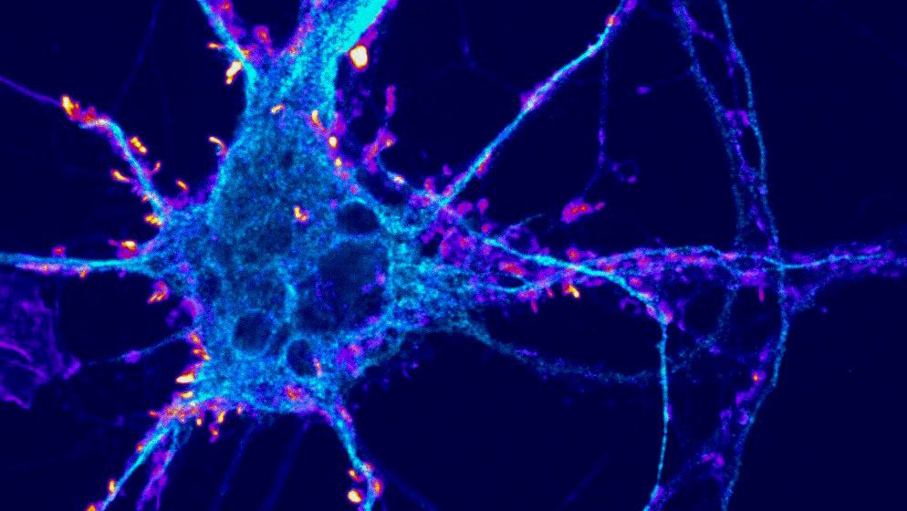
Takeaways * Scientists have made progress growing human liver in the lab. * The challenge has been to direct stems cells to grow into a mature, functioning adult organ. * This study shows that stem cells can be programmed, using genetic engineering, to grow from immature cells into mature tissue. * When a tiny lab-grown liver was transplanted into mice with liver disease, it extended the lives of the sick animals.* * *Imagine if researchers could program stem cells, which have the potential to grow into all cell types in the body, so that they could generate an entire human organ. This would allow scientists to manufacture tissues for testing drugs and reduce the demand for transplant organs by having new ones grown directly from a patient’s cells. I’m a researcher working in this new field – called synthetic biology – focused on creating new biological parts and redesigning existing biological systems. In a new paper, my colleagues and I showed progress in one of the key challenges with lab-grown organs – figuring out the genes necessary to produce the variety of mature cells needed to construct a functioning liver. Induced pluripotent stem cells, a subgroup of stem cells, are capable of producing cells that can build entire organs in the human body. But they can do this job only if they receive the right quantity of growth signals at the right time from their environment. If this happens, they eventually give rise to different cell types that can assemble and mature in the form of human organs and tissues. The tissues researchers generate from pluripotent stem cells can provide a unique source for personalized medicine from transplantation to novel drug discovery. But unfortunately, synthetic tissues from stem cells are not always suitable for transplant or drug testing because they contain unwanted cells from other tissues, or lack the tissue maturity and a complete network of blood vessels necessary for bringing oxygen and nutrients needed to nurture an organ. That is why having a framework to assess whether these lab-grown cells and tissues are doing their job, and how to make them more like human organs, is critical. Inspired by this challenge, I was determined to establish a synthetic biology method to read and write, or program, tissue development. I am trying to do this using the genetic language of stem cells, similar to what is used by nature to form human organs. Tissues and organs made by genetic designsI am a researcher specializing in synthetic biology and biological engineering at the Pittsburgh Liver Research Center and McGowan Institute for Regenerative Medicine, where the goals are to use engineering approaches to analyze and build novel biological systems and solve human health problems. My lab combines synthetic biology and regenerative medicine in a new field that strives to replace, regrow or repair diseased organs or tissues. I chose to focus on growing new human livers because this organ is vital for controlling most levels of chemicals – like proteins or sugar – in the blood. The liver also breaks down harmful chemicals and metabolizes many drugs in our body. But the liver tissue is also vulnerable and can be damaged and destroyed by many diseases, such as hepatitis or fatty liver disease. There is a shortage of donor organs, which limits liver transplantation. To make synthetic organs and tissues, scientists need to be able to control stem cells so that they can form into different types of cells, such as liver cells and blood vessel cells. The goal is to mature these stem cells into miniorgans, or organoids, containing blood vessels and the correct adult cell types that would be found in a natural organ. One way to orchestrate maturation of synthetic tissues is to determine the list of genes needed to induce a group of stem cells to grow, mature and evolve into a complete and functioning organ. To derive this list I worked with Patrick Cahan and Samira Kiani to first use computational analysis to identify genes involved in transforming a group of stem cells into a mature functioning liver. Then our team led by two of my students – Jeremy Velazquez and Ryan LeGraw – used genetic engineering to alter specific genes we had identified and used them to help build and mature human liver tissues from stem cells. The tissue is grown from a layer of genetically engineered stem cells in a petri dish. The function of genetic programs together with nutrients is to orchestrate formation of liver organoids over the course of 15 to 17 days. Liver in a dishI and my colleagues first compared the active genes in fetal liver organoids we had grown in the lab with those in adult human livers using a computational analysis to get a list of genes needed for driving fetal liver organoids to mature into adult organs. We then used genetic engineering to tweak genes – and the resulting proteins – that the stem cells needed to mature further toward an adult liver. In the course of about 17 days we generated tiny – several millimeters in width – but more mature liver tissues with a range of cells typically found in livers in the third trimester of human pregnancies. Like a mature human liver, these synthetic livers were able to store, synthesize and metabolize nutrients. Though our lab-grown livers were small, we are hopeful that we can scale them up in the future. While they share many similar features with adult livers, they aren’t perfect and our team still has work to do. For example, we still need to improve the capacity of the liver tissue to metabolize a variety of drugs. We also need to make it safer and more efficacious for eventual application in humans.[Deep knowledge, daily. Sign up for The Conversation’s newsletter.]Our study demonstrates the ability of these lab livers to mature and develop a functional network of blood vessels in just two and a half weeks. We believe this approach can pave the path for the manufacture of other organs with vasculature via genetic programming. The liver organoids provide several key features of an adult human liver such as production of key blood proteins and regulation of bile – a chemical important for digestion of food. When we implanted the lab-grown liver tissues into mice suffering from liver disease, it increased the life span. We named our organoids “designer organoids,” as they are generated via a genetic design. This article is republished from The Conversation, a nonprofit news site dedicated to sharing ideas from academic experts. It was written by: Mo Ebrahimkhani, University of Pittsburgh. Read more: * Brain organoids help neuroscientists understand brain development, but aren’t perfect matches for real brains * Why are scientists trying to manufacture organs in space?Mo Ebrahimkhani receives funding from National Institute of Health, University of Pittsburgh and Arizona Biomedical Research Council.
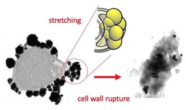
Finding alternatives to antibiotics is one of the biggest challenges facing the research community. Bacteria are increasingly resistant to these drugs, and this resistance leads to the deaths of more than 25,000 around the world. Now, a multidisciplinary team of researchers from the Universitat Rovira i Virgili, the University of Grenoble (France), the University of Saarland (Germany) and RMIT University (Australia) have discovered that the mechanical deformation of bacteria is a toxic mechanism that can kill bacteria with gold nanoparticles. The results of this research have been published in the journal Advanced Materials and are a breakthrough in researchers’ understanding the antibacterial effects of nanoparticles and their efforts to find new materials with bactericide properties.

Since the times of Ancient Egypt, gold has been used in a range of medical applications and, more recently, as for diagnosing and treating diseases such as cancer. This is due to the fact that gold is a chemically inert material, that is, it does not react or change when it comes into contact with an organism. Amongst the scientific community, nanoparticles are known for their ability to make tumors visible and for their applications in nanomedicine.

This new research shows that these chemically inert nanoparticles can kill thanks to a physical mechanism that deforms the cell wall. To demonstrate this, the researchers have synthesized in the laboratory in the shape of an almost perfect sphere and others in the shape of stars, all measuring 100 nanometres (8 times thinner than a hair). The group analyzed how these particle interact with living bacteria. “We find that the bacteria become deformed and deflate like a ball that is having the air let out before dying in the presence of these nanoparticles,” explained Vladimir Baulin, researcher at the Department of Chemical Engineering of the URV. The researchers state the bacteria seem to have died after a massive leak, “as if the cell wall had spontaneously exploded.”

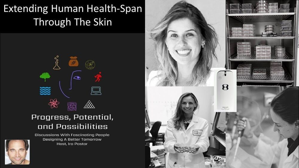
Dr. Carolina Reis Oliveria, is the CEO and Co-Founder of OneSkin Technologies, a biotechnology platform dedicated to exploring longevity science.

Carolina holds her Ph.D. in Immunology at the Federal University of Minas Gerais, in collaboration with the Rutgers University, where she conducted research with pluripotent stem cells as a source of retinal pigmented epithelium (RPE) cells, as well as the potential of RPE-stem cells derived as toxicological models for screening of new drugs with intra-ocular applications.

She founded a company called CELLSEQ solutions in Brazil which develops tools to revolutionize the safety and toxicology assays performed by pharmaceutical, cosmetic, agro-chemical and food industries, with technology based on stem cells and big data analysis.

She is an alumnus of IndieBio, the world’s leading biotechnology accelerator.

In 2016, Carolina relocated to Silicon Valley from Latin America to co-found OneSkin, and to lead the development of the company’s technologies.

“Researchers report today that they’ve created a nontoxic and nonhallucinogenic chemical cousin of ibogaine that combats depression and addictive behaviors in rodents. The work provides new hope that chemists may one day be able to create medicines for people that offer the purported therapeutic benefits of ibogaine and other psychoactive compounds without their side effects.”


Analog of ibogaine could hold hope for humans.

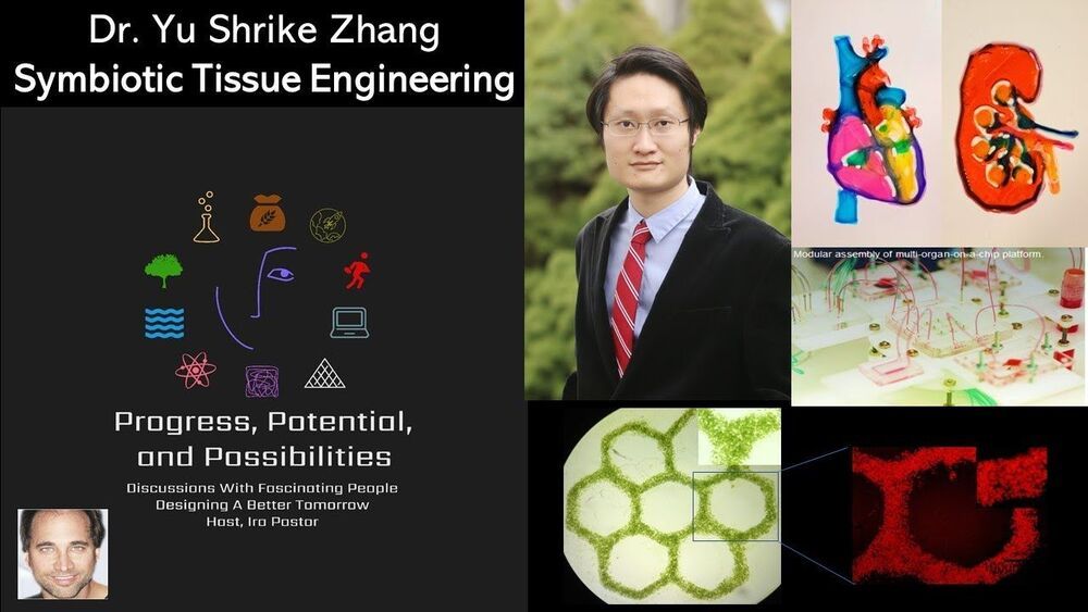
Dr yu shrike zhang phd is assistant professor at harvard medical school and associate bioengineer at brigham and women’s hospital.

Dr. Zhang’s research interests include symbiotic tissue engineering, 3D bio-printing, organ-on-a-chip technology, biomaterials, regenerative engineering, bioanalysis, nanomedicine, and biology.

His scientific contributions have been recognized by over 40 regional, national and international awards. He has been invited to deliver more than 110 lectures worldwide, and has served as reviewer for more than 500 manuscripts for as many as 50 journals.

Dr. Zhang is serving as Editor-in-Chief for Microphysiological Systems, and is Associate Editor for Bio-Design and Manufacturing, Nano Select, Aggregate, and Essays in Biochemistry.

He is also on the Editorial Board of Biofabrication, Bioprinting, Advanced Healthcare Materials, Discover Materials, BMC Biomedical Engineering, Materials Today Bio, and Chinese Chemical Letters, the Editorial Advisory Board of Heliyon and Biomicrofluidics, the International Advisory Board of Advanced NanoBiomed Research and Advanced Materials Technologies, and the Advisory Panel of Nanotechnology.

Dr. Zhang has his PhD in Biomedical Engineering from Georgia Institute of Technology / Emory, his M.S. in Bioengineering and Biomedical Engineering from Washington University in St. Louis, and his B.Eng. in Biomedical Engineering Southeast University in China.

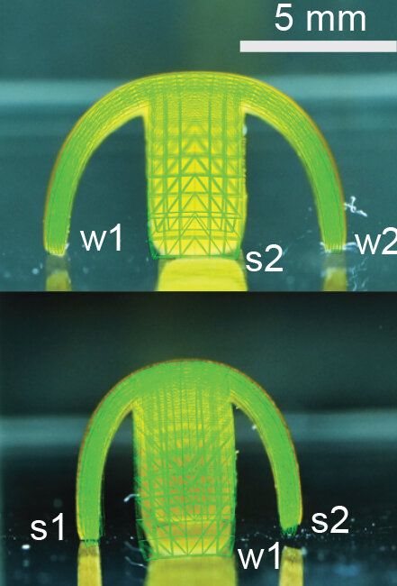
A new robot created by researchers at Northwestern University looks and behaves like a tiny aquatic animal, and could serve a variety of functions, including moving things place to place, catalyzing chemical reactions, delivering therapeutics and much more. This new soft robot honestly looks a heck of a lot like a lemon peel, but it’s actually a material made up of 90% water for the soft exterior, with a nickel skeleton inside that can change its shape in response to outside magnetic fields.

These robots are very small — only around the size of a dime — but they’re able to perform a range of tasks, including walking at the same speed as an average human, and picking up and carrying things. They work by either taking in or expelling water through their soft components, and can respond to light and magnetic fields thanks to their precise molecular design. Essentially, their molecular structure is crafted such that when they’re hit by light, the molecules that make them up expel water, causing the robot’s “legs” to stiffen like muscles.

Northwestern University researchers have developed a first-of-its-kind life-like material that acts as a soft robot. It can walk at human speed, pick up and transport cargo to a new location, climb up hills and even break-dance to release a particle.

Nearly 90% water by weight, the centimeter-sized moves without complex hardware, hydraulics or electricity. Instead, it is activated by light and walks in the direction of an external rotating .

Resembling a four-legged octopus, the robot functions inside a water-filled tank, making it ideal for use in aquatic environments. The researchers imagine customizing the movements of miniature robots to help catalyze different chemical reactions and then pump out the valuable products. The robots also could be molecularly designed to recognize and actively remove unwanted particles in specific environments, or to use their mechanical movements and locomotion to precisely deliver bio-therapeutics or cells to specific tissues.

JILA researchers have developed tools to “turn on” quantum gases of ultracold molecules, gaining control of long-distance molecular interactions for potential applications such as encoding data for quantum computing and simulations.

The new scheme for nudging a down to its lowest energy state, called quantum degeneracy, while suppressing that break up finally makes it possible to explore exotic quantum states in which all the molecules interact with one another.

The research is described in the Dec. 10 issue of Nature. JILA is a joint institute of the National Institute of Standards and Technology (NIST) and the University of Colorado Boulder.

The observation of a chemical reaction at the molecular level in real time is a central theme in experimental chemical physics. An international research team has captured roaming molecular fragments for the first time. The work, under the supervision of Heide Ibrahim, research associate at the Institut national de la recherche scientifique (INRS), was published in the journal Science.

The research group of the Énergie Matériaux Télécommunications Research Centre of INRS, with support of Professor François Légaré, has used the Advanced Laser Light Source (ALLS). They have succeeded in shooting the first molecular film of “roamers”—hydrogen fragments, in this case—that orbit around HCO fragments) during a chemical reaction by studying the photo-dissociation of formaldehyde, H2CO.

In groundbreaking new research, an international team of researchers led by the University of Minnesota Twin Cities has developed a unique process for producing a quantum state that is part light and part matter.

The discovery provides fundamental new insights for more efficiently developing the next generation of quantum-based optical and electronic devices. The research could also have an impact on increasing efficiency of nanoscale chemical reactions.

The research is published in Nature Photonics.