MIT has launched the Stephen A. Schwarzman College of Computing, a $1 billion center dedicated to “reshaping its academic program” around AI. The idea, said MIT president L. Rafael Reif, is to use AI, machine learning and data science with other academic disciplines to “educate the bilinguals of the future,” defining bilingual as those working in biology, chemistry, politics, history and linguistics with computing skills that can be used in their field.
Category: biological
What happens when a new technology is so precise that it operates on a scale beyond our characterization capabilities? For example, the lasers used at INRS produce ultrashort pulses in the femtosecond range (10-15 s), which is far too short to visualize. Although some measurements are possible, nothing beats a clear image, says INRS professor and ultrafast imaging specialist Jinyang Liang. He and his colleagues, led by Caltech’s Lihong Wang, have developed what they call T-CUP: the world’s fastest camera, capable of capturing 10 trillion (1013) frames per second (Fig. 1). This new camera literally makes it possible to freeze time to see phenomena—and even light—in extremely slow motion.
In recent years, the junction between innovations in non-linear optics and imaging has opened the door for new and highly efficient methods for microscopic analysis of dynamic phenomena in biology and physics. But harnessing the potential of these methods requires a way to record images in real time at a very short temporal resolution—in a single exposure.
Using current imaging techniques, measurements taken with ultrashort laser pulses must be repeated many times, which is appropriate for some types of inert samples, but impossible for other more fragile ones. For example, laser-engraved glass can tolerate only a single laser pulse, leaving less than a picosecond to capture the results. In such a case, the imaging technique must be able to capture the entire process in real time.
Can the origin of life be explained with quantum mechanics? And if so, are there quantum algorithms that could encode life itself?
We’re a little closer to finding out the answers to those big questions thanks to new research carried out with an IBM supercomputer.
Encoding behaviours related to self-replication, mutation, interaction between individuals, and (inevitably) death, a newly created quantum algorithm has been used to show that quantum computers can indeed mimic some of the patterns of biology in the real world.
https://www.youtube.com/watch?v=n4YtQ-N_t84
When listening to world science festival’s latest episode on youtube, Pondering the Imponderables: The Biggest Questions of Cosmology, I found myself to be most in line with George F.R. Ellis’ line of thinking overall.
Big Bang cosmology, chemical and biological evolutionary theory, and associated sciences have been extraordinarily successful in revealing and enabling us to understand the development of the.
Cosmology is today a precision science with masses of high quality data every increasing our understanding of the physical universe, but paradoxically theoretical cosmology is simultaneously.
Professor George Ellis FRS Introduced by Prof. Alister McGrath Followed by a panel discussion with Prof. Ard Louis and Prof. Denis Noble Mathematical Institute, Oxford.
Fuel cells have long been viewed as a promising power source. These devices, invented in the 1830s, generate electricity directly from chemicals, such as hydrogen and oxygen, and produce only water vapor as emissions. But most fuel cells are too expensive, inefficient, or both.
In a new approach, inspired by biology and published today (Oct. 3, 2018) in the journal Joule, a University of Wisconsin-Madison team has designed a fuel cell using cheaper materials and an organic compound that shuttles electrons and protons.
In a traditional fuel cell, the electrons and protons from hydrogen are transported from one electrode to another, where they combine with oxygen to produce water. This process converts chemical energy into electricity. To generate a meaningful amount of charge in a short enough amount of time, a catalyst is needed to accelerate the reactions.
In a new scientific article, researchers at Uppsala University describe how, using a completely new method, they have synthesised an artificial enzyme that functions in the metabolism of living cells. These enzymes can utilize the cell’s own energy, and thereby enable hydrogen gas to be produced from solar energy.
Hydrogen gas has long been noted as a promising energy carrier, but its production is still dependent on fossil raw materials. Renewable hydrogen gas can be extracted from water, but as yet the systems for doing so have limitations.
In the new article, published in the journal Energy and Environmental Science, an interdisciplinary European research group led by Uppsala University scientists describe how artificial enzymes convert solar energy into hydrogen gas. This entirely new method has been developed at the University in the past few years. The technique is based on photosynthetic microorganisms with genetically inserted enzymes that are combined with synthetic compounds produced in the laboratory. Synthetic biology has been combined with synthetic chemistry to design and create custom artificial enzymes inside living organisms.
A fluorescent molecule whose luminosity depends upon how fast it can rotate is helping researchers measure how viscous the fluid is inside different parts of a cell.
“There’s a lot of interest in the biophysical field in developing chemical probes that can be used to characterize the environment inside a cell or any kind of biological compartment,” says Peter Bond, from A*STAR’s Bioinformatics Institute.
Researchers from the United Kingdom and Singapore—including A*STAR scientists such as Bond’s team who led the computational arm of the project—have modeled, developed and tested a molecule comprising two parts; a genetic probe designed to home in on particular proteins, so it can be directed to wherever in a cell that protein is found; and a molecular rotor—a fluorescent molecule whose fluorescence lasts longer, the slower it spins. A*STAR researchers simulated how this molecule would perform in different microenvironments at scales of millionths or even billionths of a meter.
👀
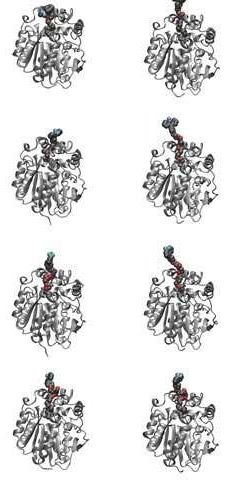
The GTPases constitute a very large protein family, whose members are involved in the control of cell growth, transport of molecules, synthesis of other proteins, etc. Despite the many functions of the GTPases, they follow a common cyclic pattern (Figure 1). The activity of the GTPases is regulated by factors that control their ability to bind and hydrolyse guanosine triphosphate (GTP) to guanosine diphosphate (GDP). So far, it has been the general assumption that a GTPase is active or “on” when it is bound to GTP and inactive or “off” in complex with GDP. The GTPases are therefore sometimes referred to as molecular “switches.”
The bacterial translational elongation factor EF-Tu is a GTPase, which plays a crucial role during the synthesis of proteins in bacteria, as the factor transports the amino acids that build up a cell’s proteins to the cellular protein synthesis factory, the ribosome. Previous structural studies using X-ray crystallography have shown that EF-Tu occurs in two markedly different three-dimensional shapes depending on whether the factor is “on” (i.e. bound to GTP) or “off” (i.e. bound to GDP) (Figure 2). The binding of GTP/GDP have therefore always been thought to be decisive for the factor’s structural conformation.
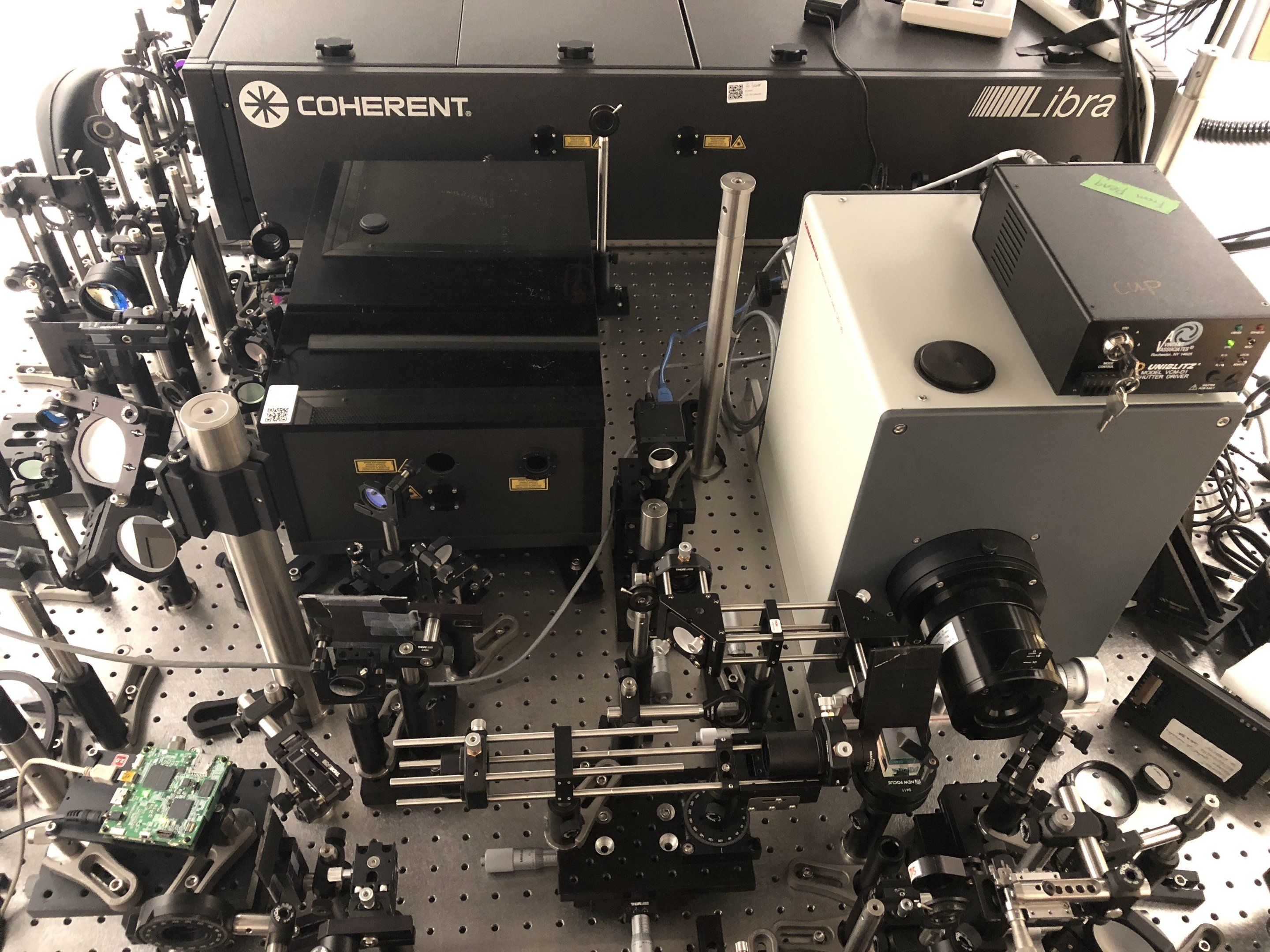
However, a research collaboration between researchers from the Department of Molecular Biology and Genetics at Aarhus University and two American universities reveals that EF-Tu’s structure and function, and probably also those of other GTPases, are far more complex than previously assumed. In Søren Thirup’s group, X-ray crystallographic analysis of E. coli EF-Tu has shown that EF-Tu bound to a variant of GTP, GDPNP, can also occur in the “off” state, which is characterised by a more open structure. In collaboration with American researchers, Charlotte Knudsen’s Ph.D. student, Darius Kavaliauskas, conducted further studies using a special form of fluorescence microscopy that makes it possible to observe the spatial structure of individual EF-Tu molecules in solution.